Copyright
©The Author(s) 2020.
World J Clin Cases. Jun 26, 2020; 8(12): 2623-2628
Published online Jun 26, 2020. doi: 10.12998/wjcc.v8.i12.2623
Published online Jun 26, 2020. doi: 10.12998/wjcc.v8.i12.2623
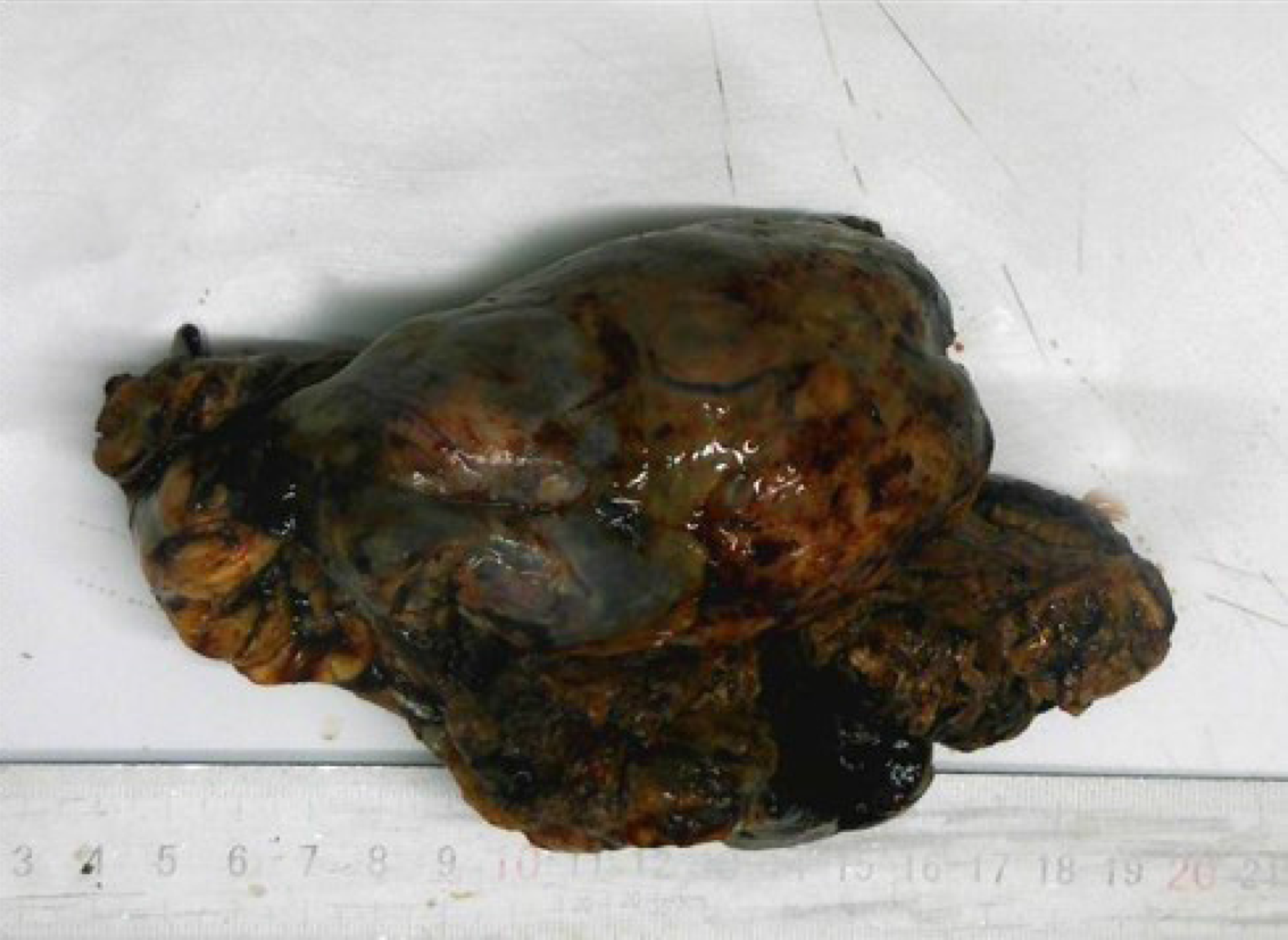
Figure 1 The mass was dark brown, and the cut surface was solid, thick-nodular, tender in texture, and grayish and gray-yellow in color.
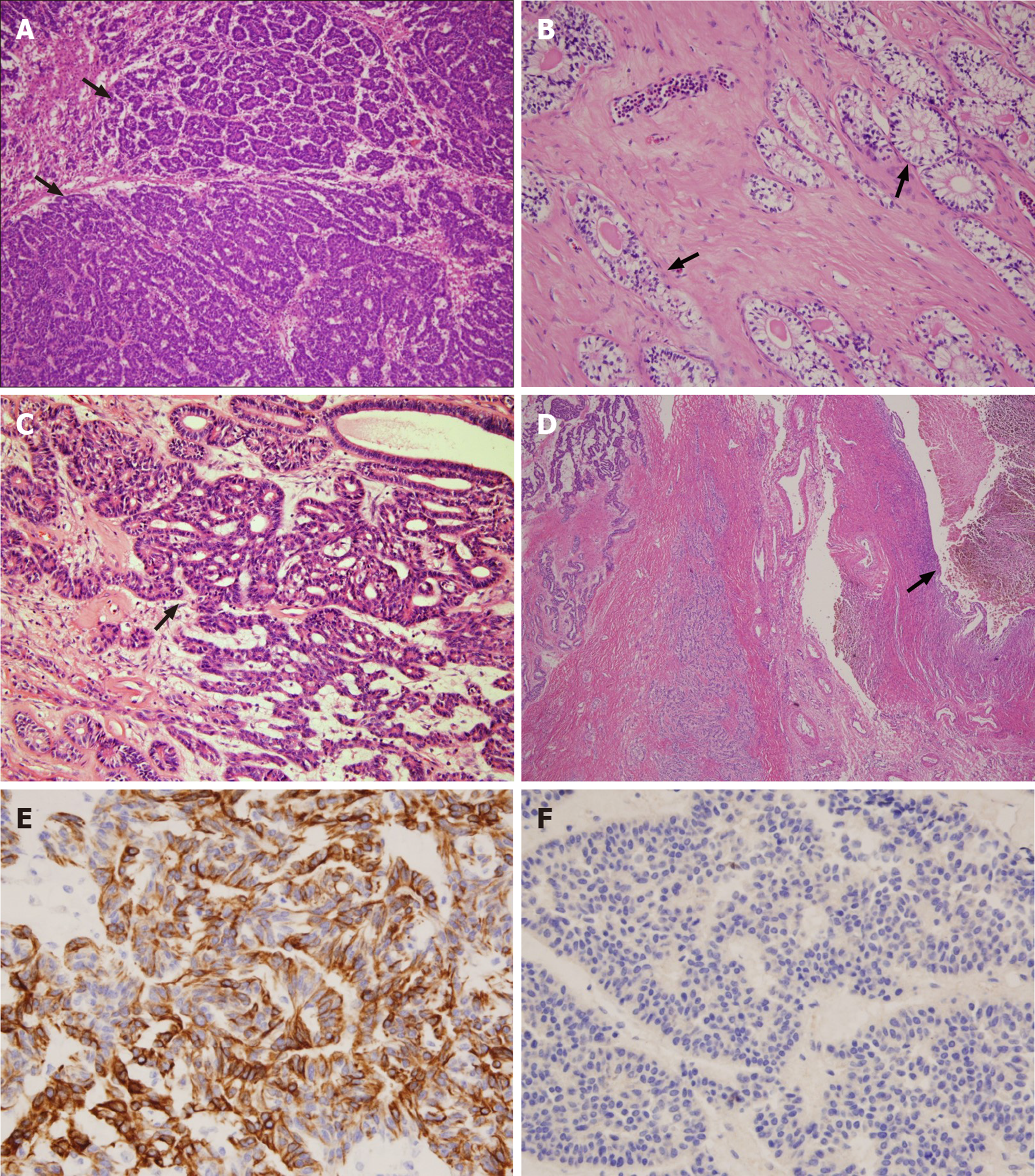
Figure 2 Pathological examination (HE staining) of the tumor and immunohistochemical analysis.
A: The tumor consisted of micro-adenoids, trabeculae, and small tubular structures that resembled granulosa cell tumor (black arrow; HE, × 100); B: Small tubular structures that resembled sertoli cell tumor (black arrow; HE, × 200); C: Areas of typical endometrioid carcinoma (black arrow; HE, × 200); D: Endometriotic cyst was seen next to the tumor (black arrow; HE, × 40); E: Tumor cells diffusely expressed CK7 (EnVision, × 400); F: Tumor cells did not express α-inhibin (EnVision, × 400).
- Citation: Wei XX, He YM, Jiang W, Li L. Ovarian endometrioid carcinoma resembling sex cord-stromal tumor: A case report. World J Clin Cases 2020; 8(12): 2623-2628
- URL: https://www.wjgnet.com/2307-8960/full/v8/i12/2623.htm
- DOI: https://dx.doi.org/10.12998/wjcc.v8.i12.2623


