Copyright
©The Author(s) 2020.
World J Clin Cases. May 26, 2020; 8(10): 2038-2043
Published online May 26, 2020. doi: 10.12998/wjcc.v8.i10.2038
Published online May 26, 2020. doi: 10.12998/wjcc.v8.i10.2038
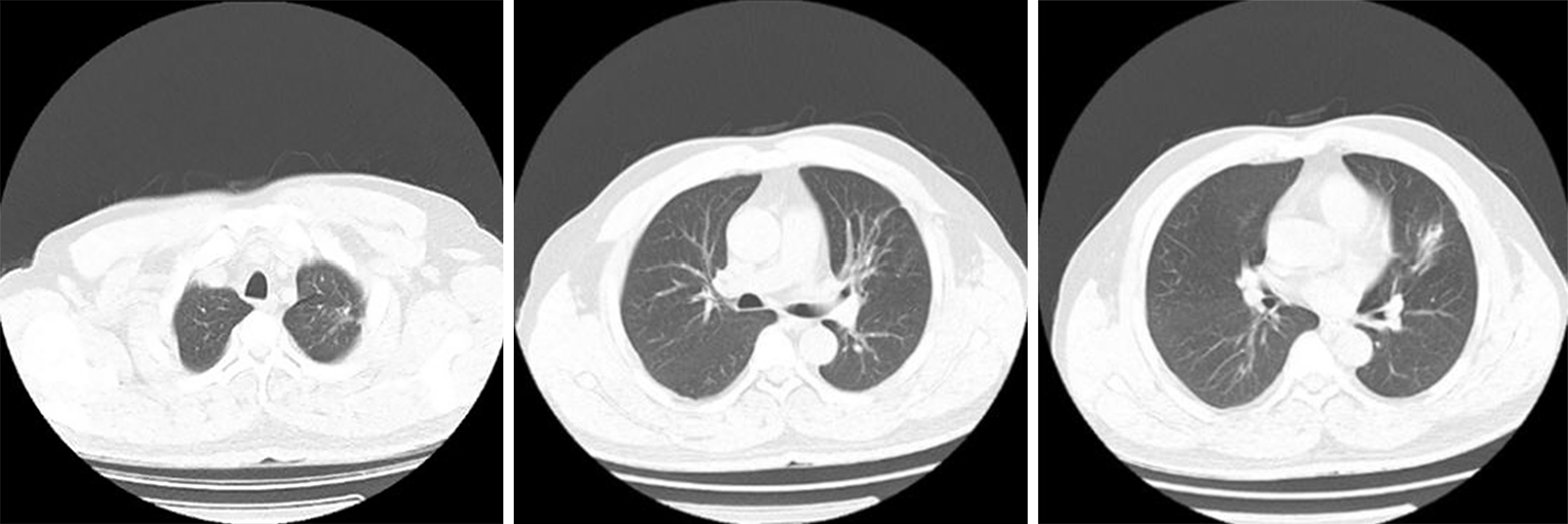
Figure 1 Multiple modes and areas of patchy increased density were evident in the upper left lung.
Figure 2 Hematoxylin and eosin and periodic-acid-Schiff-stained lung tissue sections highlighted the presence of granulomatous inflammation containing yeast-like microbes that were surrounded by clear halos within multinucleated giant cells and in intercellular spaces.
Figure 3 Multiple modes and areas of patchy increased density were evident in the upper left lung, with no significant changes relative to Figure 1.
Figure 4 Multiple modes and areas of patchy increased density were evident in the upper left lung, with significant reductions relative to Figure 3.
Figure 5 A small cable-like area of increased density was evident in the upper left lung, consistent with a > 90% reduction in the mass relative to the previous examination.
- Citation: Jiang XQ, Zhang YB. Cryptococcal pneumonia in a human immunodeficiency virus-negative patient: A case report. World J Clin Cases 2020; 8(10): 2038-2043
- URL: https://www.wjgnet.com/2307-8960/full/v8/i10/2038.htm
- DOI: https://dx.doi.org/10.12998/wjcc.v8.i10.2038


