Copyright
©The Author(s) 2019.
World J Clin Cases. Apr 6, 2019; 7(7): 891-897
Published online Apr 6, 2019. doi: 10.12998/wjcc.v7.i7.891
Published online Apr 6, 2019. doi: 10.12998/wjcc.v7.i7.891
Figure 1 Pathological analysis of the surgical specimen (hematoxylin and eosin stain, original magnification, 40 ×).
A: The tumor was composed of blunt finger-like processes lined by pseudostratified columnar cells with frequent goblet cells (arrow); B: Intracellular and extracellular abundant mucin was seen (arrow); C: The carcinomatous areas consisted of dysplastic glands and some of the glands presented with high-grade intraepithelial neoplasia (arrow).
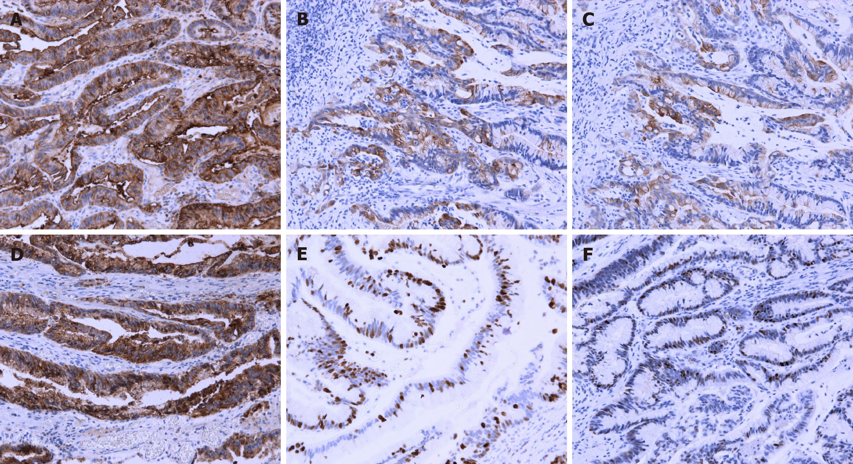
Figure 2 Immunohistochemical analysis of the surgical specimen (magnification, 200 ×).
A: Carcinoembryonic antigen was stained positive; B: Cytokeratin 7 was stained positive; C: Cytokeratin 20 was stained positive; D: Epithelial membrane antigen was stained positive; E: The positive ratio of Ki-67 was 60%; F: p53 was stained positive.
- Citation: Qin LF, Liang Y, Xing XM, Wu H, Yang XC, Niu HT. Villous adenoma coexistent with focal well-differentiated adenocarcinoma of female urethral orifice: A case report and review of literature. World J Clin Cases 2019; 7(7): 891-897
- URL: https://www.wjgnet.com/2307-8960/full/v7/i7/891.htm
- DOI: https://dx.doi.org/10.12998/wjcc.v7.i7.891


