Copyright
©The Author(s) 2019.
World J Clin Cases. Oct 26, 2019; 7(20): 3364-3371
Published online Oct 26, 2019. doi: 10.12998/wjcc.v7.i20.3364
Published online Oct 26, 2019. doi: 10.12998/wjcc.v7.i20.3364
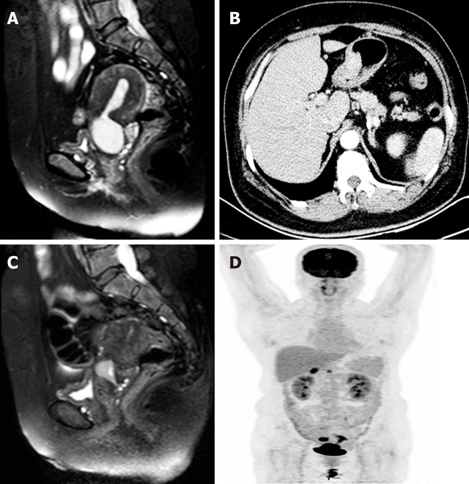
Figure 1 Imaging findings of the patient.
A: Pre-chemotherapy magnetic resonance imaging (MRI) scan showed a solid mass located in cervical canal of uterus, suspected involvement of rectal wall; B: Thicken gastric wall was suspected malignant on computed tomography (CT) scan; C: Postchemotherapy MRI scan showed a decrescent solid mass located in cervical canal of uterus; D: Abnormal fluorodeoxyglucose uptake in the cervix uterus, uterine cavity, right adnexa as well as in the stomach on positron emission topography/CT scan.
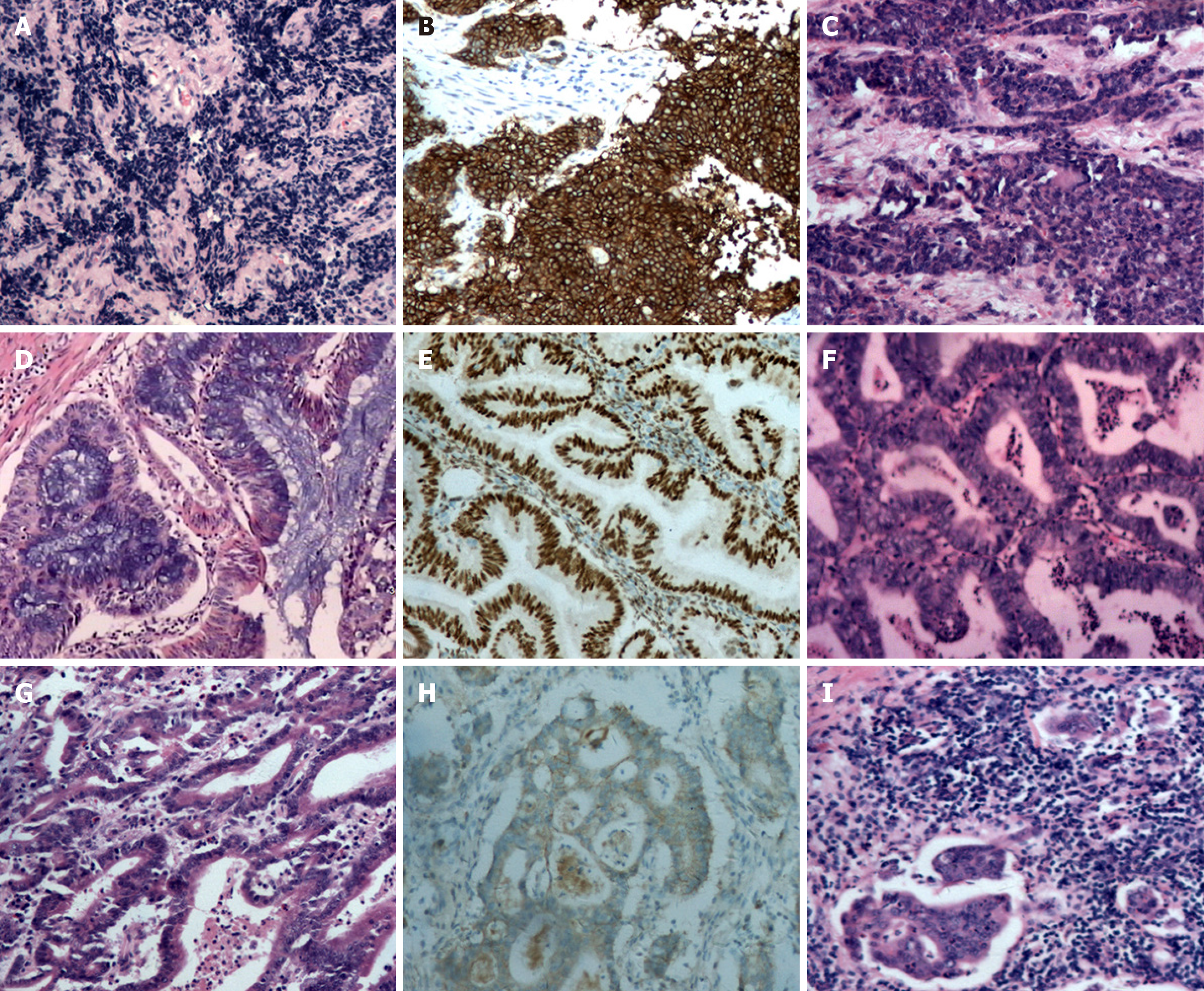
Figure 2 Histopathological and immunohistochemical staining findings.
A: Poorly differentiated endocervical adenocarcinoma; B: Positive stain for Syn; C: Metastatic adenocarcinoma of rectal wall; D: Diffuse atypical hyperplasia in endometrium with focal highly differentiated endometroid adenocarcinoma; E: Positive stain for ER; F: Endometroid adenocarcinoma in right ovary; G: Highly to moderately differentiated adenocarcinoma in stomach; H: Positive stain for Her-2; I: Metastatic adenocarcinoma in perigastric lymph node.
- Citation: Wang DD, Yang Q. Synchronous quadruple primary malignancies of the cervix, endometrium, ovary, and stomach in a single patient: A case report and review of literature. World J Clin Cases 2019; 7(20): 3364-3371
- URL: https://www.wjgnet.com/2307-8960/full/v7/i20/3364.htm
- DOI: https://dx.doi.org/10.12998/wjcc.v7.i20.3364


