Copyright
©The Author(s) 2024.
World J Clin Cases. Feb 16, 2024; 12(5): 931-941
Published online Feb 16, 2024. doi: 10.12998/wjcc.v12.i5.931
Published online Feb 16, 2024. doi: 10.12998/wjcc.v12.i5.931
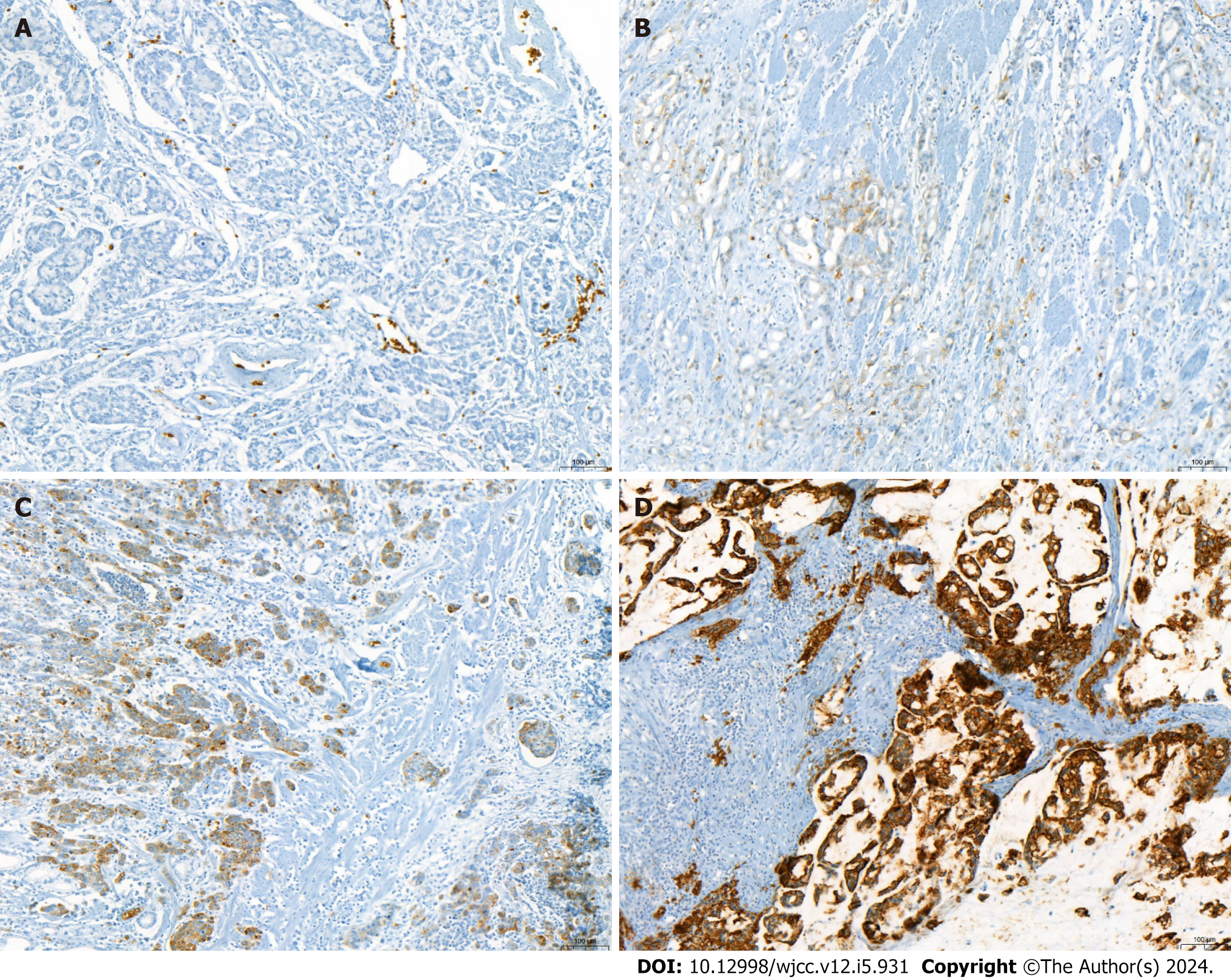
Figure 1 Immunohistochemical staining for glucose transport protein 1 in colorectal cancer.
Glucose transport protein 1 expression was demonstrated as cytoplasmic or membranous staining. A-D: The score was assessed according to the intensity [0 = negative (A), 1 = weak (B), 2 = moderate (C), 3 = strong (D); × 100]. A score of 2 or higher was considered as positive.
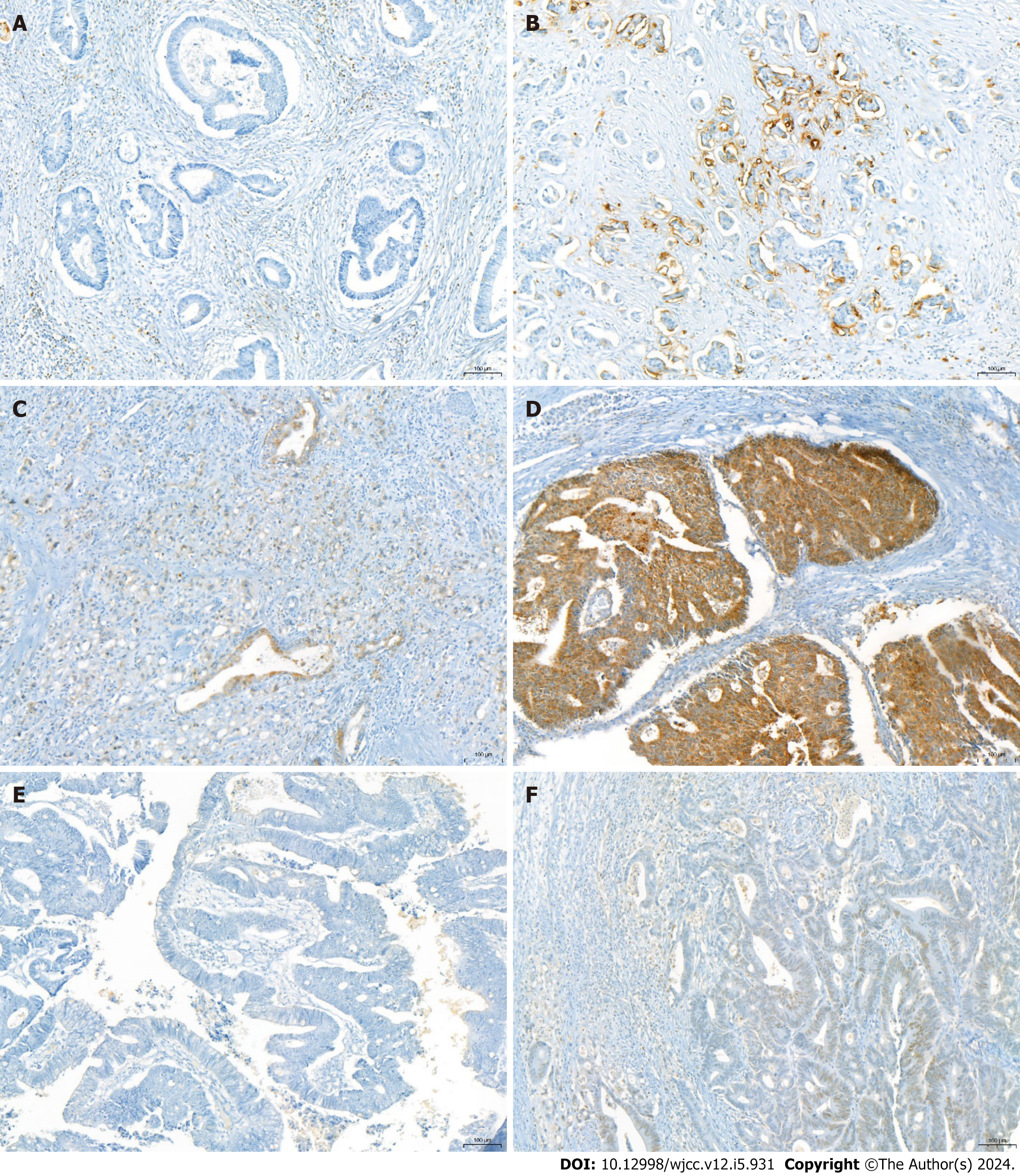
Figure 2 Immunohistochemical staining for glucose transport protein 3, hexokinase-II, and hypoxia-inducible factor-1 in colorectal cancer.
Glucose transport protein 3 (GLUT-3) and hexokinase-II expressions were demonstrated as cytoplasmic or membranous staining. A: GLUT-3 negative; B: GLUT-3 positive; C: Hexokinase-II negative; D: Hexokinase-II positive; E and F: Hypoxia-inducible factor-1 (HIF-1) expression was demonstrated as nuclear staining (E: HIF-1 negative, F: HIF-1 positive).
- Citation: Kim H, Choi SY, Heo TY, Kim KR, Lee J, Yoo MY, Lee TG, Han JH. Value of glucose transport protein 1 expression in detecting lymph node metastasis in patients with colorectal cancer. World J Clin Cases 2024; 12(5): 931-941
- URL: https://www.wjgnet.com/2307-8960/full/v12/i5/931.htm
- DOI: https://dx.doi.org/10.12998/wjcc.v12.i5.931


