Copyright
©The Author(s) 2024.
World J Clin Cases. Aug 6, 2024; 12(22): 5124-5130
Published online Aug 6, 2024. doi: 10.12998/wjcc.v12.i22.5124
Published online Aug 6, 2024. doi: 10.12998/wjcc.v12.i22.5124
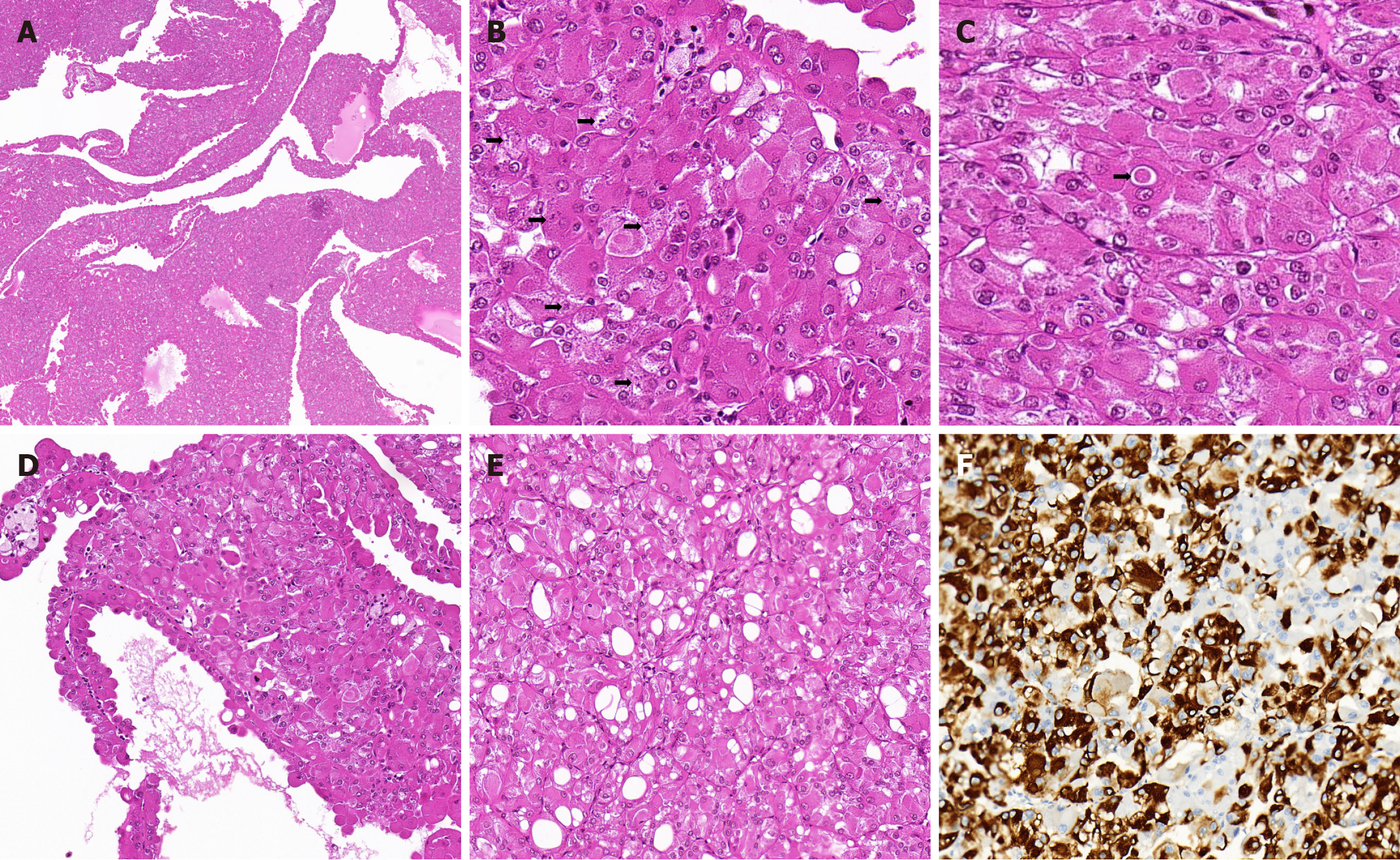
Figure 1 Images of hematoxylin and eosin staining and immunostaining in case 1.
A: Low-power microscopic examination revealed eosinophilic solid and cystic renal cell carcinoma (ESC RCC) with cystic and solid architecture; B: The tumor cells had abundant granular eosinophilic cytoplasm with prominent fine or coarse basophilic granular cytoplasmic stippling; C: Eosinophilic cytoplasmic globules; D: The tumor cells lining the septa showed a hobnail arrangement; E: Focal intracytoplasmic vacuoles were observed; F: CK20 expression in ESC RCC cells.
Figure 2 Images of hematoxylin and eosin staining and immunostaining in case 2.
A: Tumor cells were arranged in an acinar-like, microcyst, and pseudo-papillary pattern, and cells lining septa had a hobnail morphology; B: The cells exhibited a voluminous eosinophilic cytoplasm with various sizes of basophilic granular stippling; C: Immunohistochemical staining for CK20.
- Citation: Cao HH, Li H, Guo XH, Cao ZX, Zhang BH. Eosinophilic solid and cystic renal cell carcinoma with aggressive behavior: Two case reports. World J Clin Cases 2024; 12(22): 5124-5130
- URL: https://www.wjgnet.com/2307-8960/full/v12/i22/5124.htm
- DOI: https://dx.doi.org/10.12998/wjcc.v12.i22.5124


