Copyright
©The Author(s) 2024.
World J Clin Cases. Jul 6, 2024; 12(19): 3985-3994
Published online Jul 6, 2024. doi: 10.12998/wjcc.v12.i19.3985
Published online Jul 6, 2024. doi: 10.12998/wjcc.v12.i19.3985
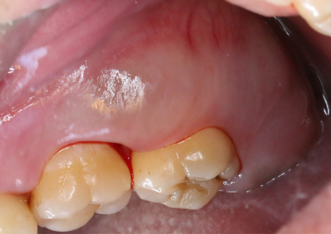
Figure 1
Initial intra-oral examination demonstrated swelling in the left maxillary molar buccal gingival region.
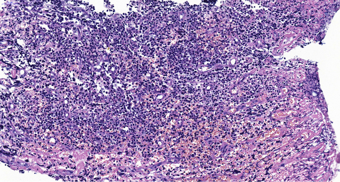
Figure 2 A photomicrograph demonstrated the presence of diffuse neoplastic infiltration in the gingiva, indicating the involvement of abnormal cells that invaded the tissue in a scattered manner.
These neoplastic cells are described as intermediate-sized immature blast-like cells with thin cytoplasm, finely-dispersed chromatin, and rounded or indented nuclei. Magnification, × 300.
Figure 3 Neoplastic cells stained positive for myeloperoxidase-positive and Ki-67.
Magnification, × 200. MPO: Myeloperoxidase-positive.
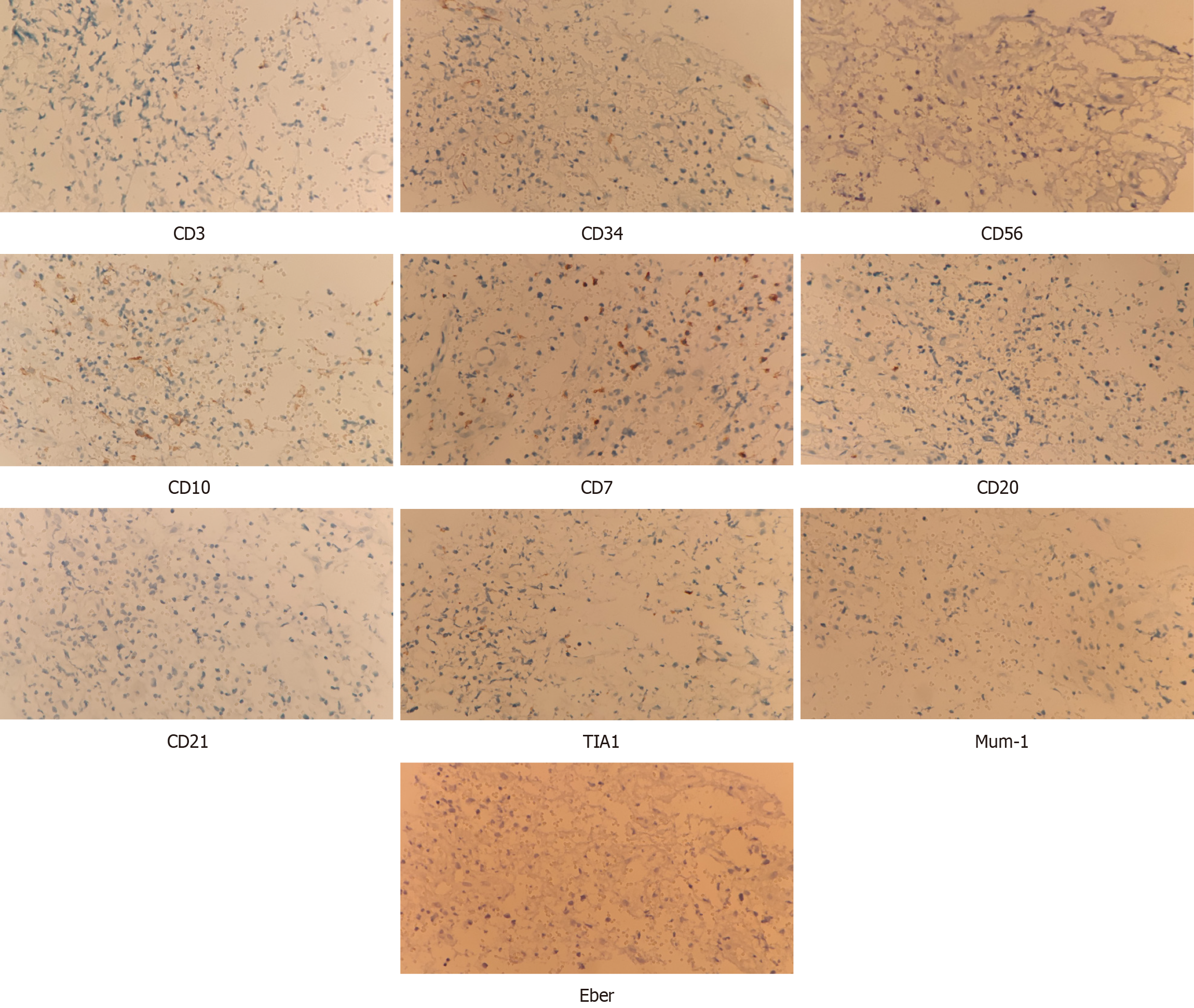
Figure 4 Cells were negative for CD3, CD34, CD56, CD10, CD7, CD20, CD21, TIA1, Mum-1 and Eber.
Magnification, × 200.
Figure 5 The proportion of red blood cells and granulocytes was as expected.
A: Various stages of red blood cells; B: Various stages of granulocytes. Red cell stages were dominated by mature cells. Megakaryocytes were visible with lobulated nuclei. Few plasma cells of lymphocytes were observed. Both fe and reticular fibers were positive. Magnification, × 300.
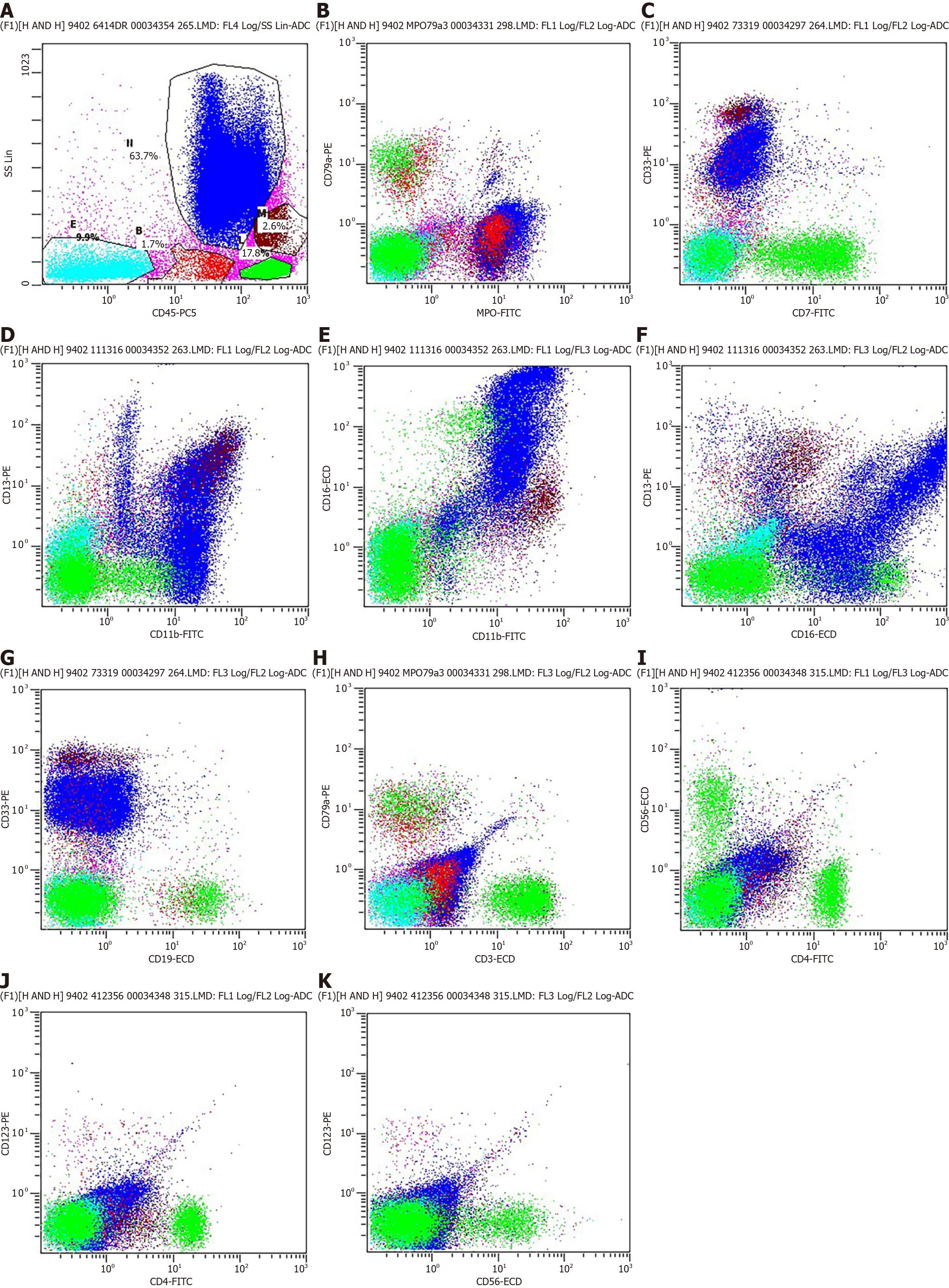
Figure 6 Immunophenotyping using flow cytometry.
A: A 63.7% of the cells were granulocytes, 17.8% of the cells were lymphocytes, 2.6% of the cells were monocytes, 1.7% of the cells were immature cells and 9.9% of the cells were CD45-negative; B-H: Flow cytometry was performed using myeloperoxidase-positive (MPO), CD79a, CD7, CD33, CD11b, CD13, CD16, CD19 and CD3 antibodies. The MPO/CD79a, CD7/CD33, CD11b/CD13, CD11b/CD16, CD16/CD13, CD19/CD33 and CD3/CD79a gate helps distinguish granulocytes morphology. The results showed that granulocytes morphology were generally within normal parameters; I-K: Flow cytometry was performed using CD4, CD56 and CD123 antibodies. The CD4/CD56, CD4/CD123 and CD56/CD123 gate helps distinguish B-cell. No distinct expressing CD4, CD56, and CD123 were observed within the B-cell group in the present study. FITC: Fluorescein isothiocyanate; ECD: Extracellular domain; ADC: Apparent diffusion coefficient.
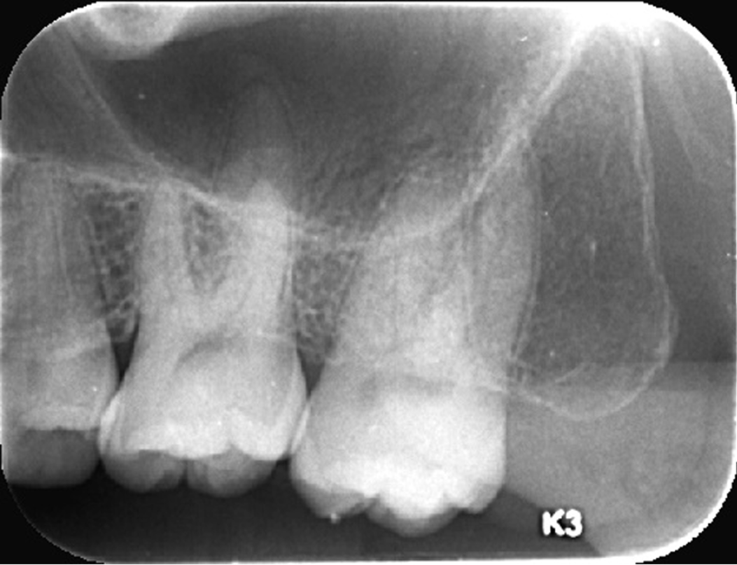
Figure 7
Radiological examination demonstrated left maxillary second molar in the absence of bone destruction.
- Citation: Li SH, Yang CX, Xing XM, Gao XR, Lu ZY, Ji QX. Myeloid sarcoma with maxillary gingival swelling as the initial symptom: A case report and review of literature. World J Clin Cases 2024; 12(19): 3985-3994
- URL: https://www.wjgnet.com/2307-8960/full/v12/i19/3985.htm
- DOI: https://dx.doi.org/10.12998/wjcc.v12.i19.3985


