Copyright
©The Author(s) 2023.
World J Clin Cases. Mar 6, 2023; 11(7): 1642-1649
Published online Mar 6, 2023. doi: 10.12998/wjcc.v11.i7.1642
Published online Mar 6, 2023. doi: 10.12998/wjcc.v11.i7.1642
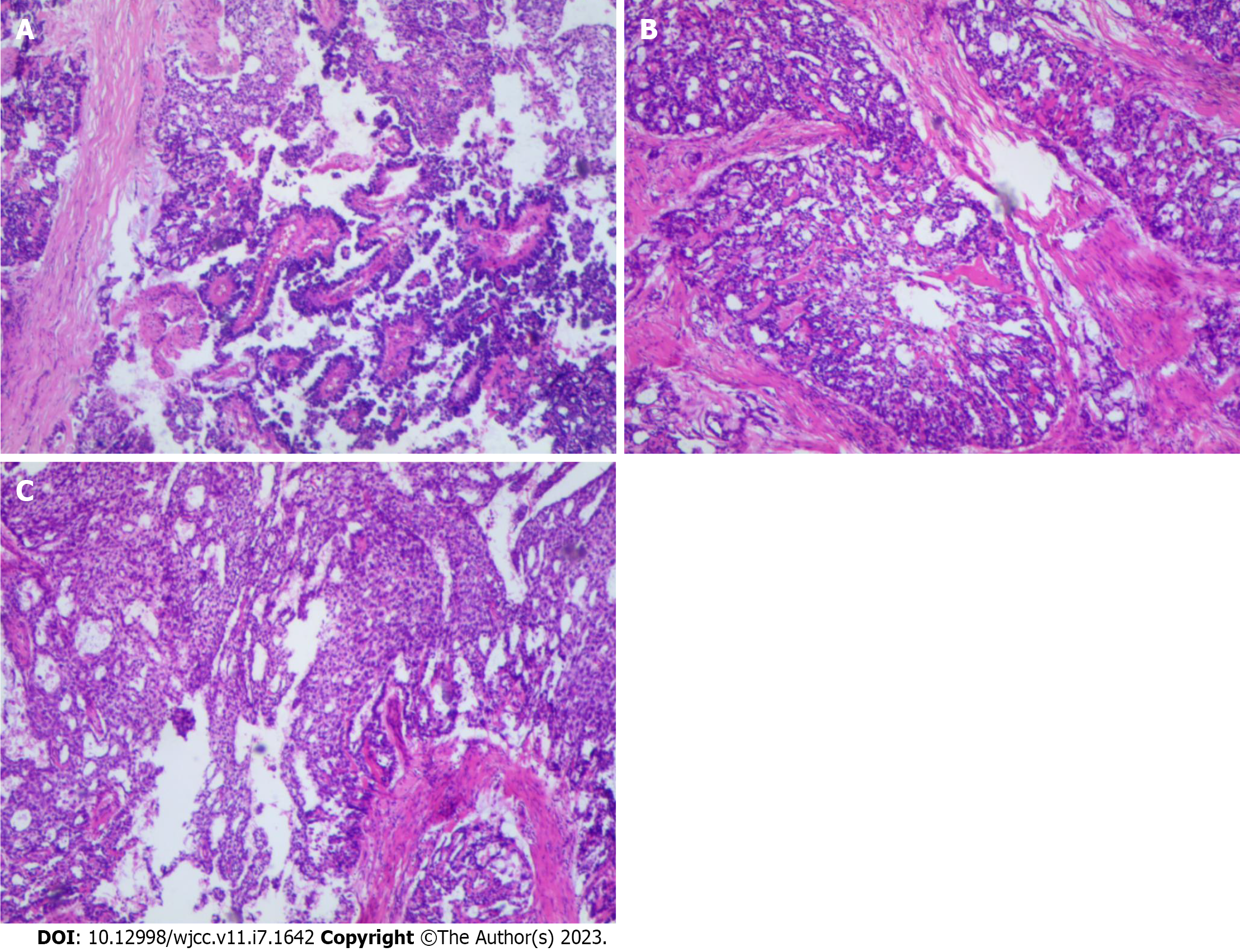
Figure 1 Histological findings from the resected tumor specimen showed at low power magnification.
A: Histological findings from the resected tumor specimen showed papillary morphology areas at low power magnification; B: Classic micro vesicular and cystically areas at low power magnification; C: A solid trabecular areas at low power magnification. Original magnification × 100 (Hematoxylin & eosin).
Figure 2 Histological findings from the resected tumor specimen showed at high power magnification.
A: Characteristic Schiller-Duval bodies; B: Loose reticular structure; C: Papillary structure; D: Solid structure; E: Mucoid structure; F: Eosinophilic globules or hyaline bodies. Original magnification × 200 (Hematoxylin & eosin).
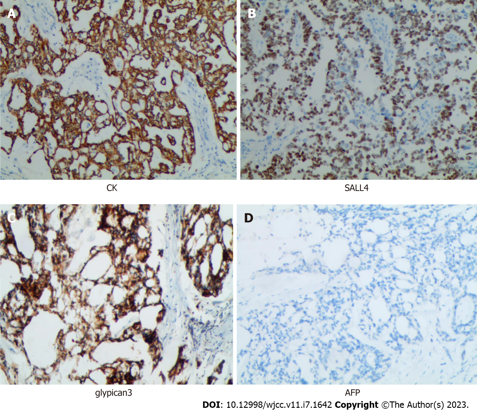
Figure 3 Immunostaining of tumor cells.
Immunostaining showed that broad-spectrum creatine kinase and glypican-3 were strongly positive in almost all the neoplastic cells; Spalt-like transcription factor 4 was strongly positive in the nuclei of the neoplastic cells; alpha-fetoprotein was negative in the neoplastic cells. Original magnification × 200 (Immunohistochemical). CK: Creatine kinase; SALL4: Spalt-like transcription factor 4; AFP: Alpha-fetoprotein.
- Citation: Wang Y, Yang J. Primary yolk sac tumor in the abdominal wall in a 20-year-old woman: A case report . World J Clin Cases 2023; 11(7): 1642-1649
- URL: https://www.wjgnet.com/2307-8960/full/v11/i7/1642.htm
- DOI: https://dx.doi.org/10.12998/wjcc.v11.i7.1642


