Copyright
©The Author(s) 2023.
World J Clin Cases. May 16, 2023; 11(14): 3351-3355
Published online May 16, 2023. doi: 10.12998/wjcc.v11.i14.3351
Published online May 16, 2023. doi: 10.12998/wjcc.v11.i14.3351
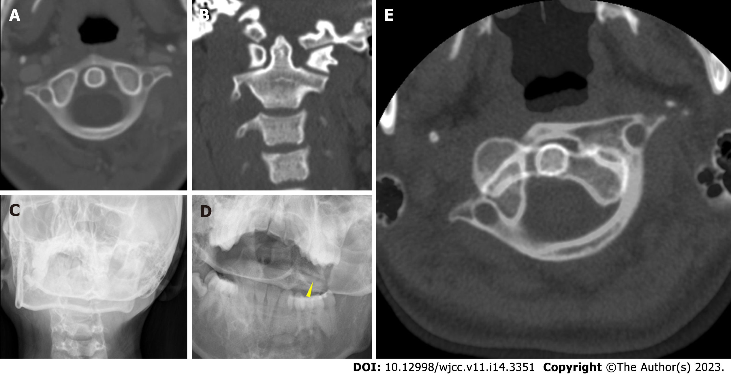
Figure 1 Preoperative imaging.
A: Computed tomography of the neck before thyroidectomy showing no subluxation of C1–2; B: Computed tomography coronal view after thyroidectomy showing the asymmetry of the lateral atlantodental interval; C: Plain radiograph showing the “cock robin” position of the head; D: Open mouth view showing the asymmetrical atlantodental interval and narrowing atlantoaxial joints (yellow arrow); E: Computed tomography showing the anterior subluxation of C1 on C2 at the left facet joint and the posterior subluxation of C1 on C2 at the right facet joint.
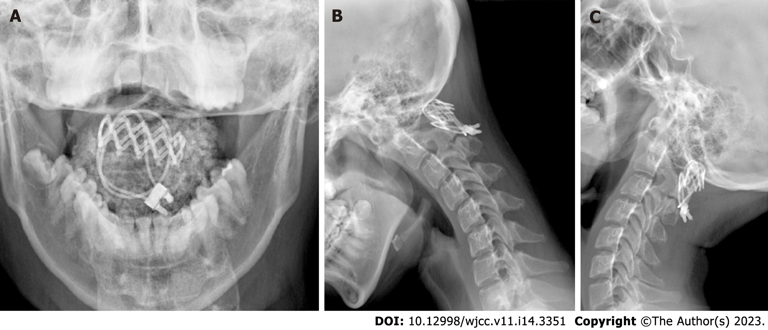
Figure 2 Postoperative cervical dynamic radiographs of a stable atlantoaxial joint.
A: Open mouth radiograph showing the reduction of the “cock robin” position of the head; B and C: Lateral flexion dynamic radiograph showing C1–2 fusion and no instability.
- Citation: Hong WJ, Lee JK, Hong JH, Han MS, Lee SS. Iatrogenic atlantoaxial rotatory subluxation after thyroidectomy in a pediatric patient: A case report. World J Clin Cases 2023; 11(14): 3351-3355
- URL: https://www.wjgnet.com/2307-8960/full/v11/i14/3351.htm
- DOI: https://dx.doi.org/10.12998/wjcc.v11.i14.3351


