Copyright
©The Author(s) 2023.
World J Clin Cases. Apr 16, 2023; 11(11): 2502-2509
Published online Apr 16, 2023. doi: 10.12998/wjcc.v11.i11.2502
Published online Apr 16, 2023. doi: 10.12998/wjcc.v11.i11.2502
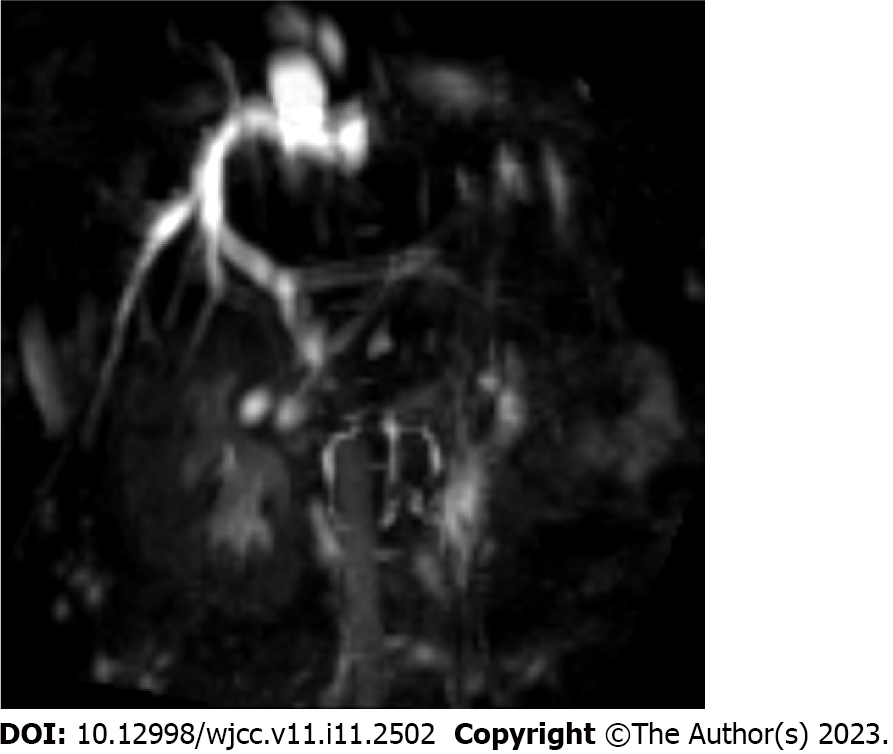
Figure 1 Magnetic resonance cholangiopancreatography examination demonstrating dilatation of the common bile duct in the porta hepatis with the widest diameter of approximately 12 mm.
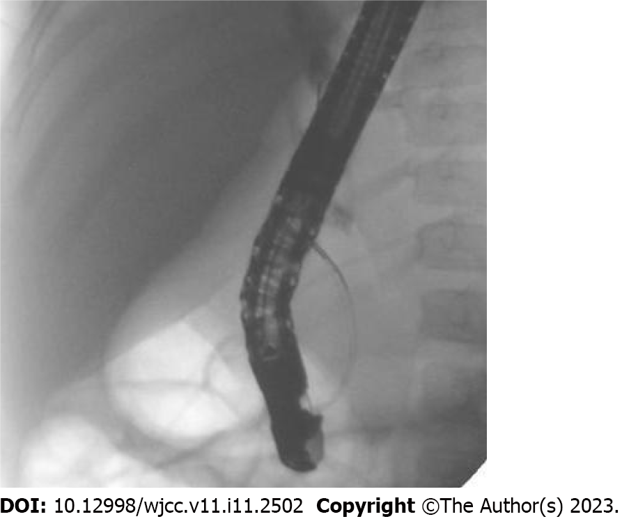
Figure 2 Aggregation of contrast in the porta hepatis and non-visualization of the intrahepatic bile duct and the middle and lower common bile ducts.
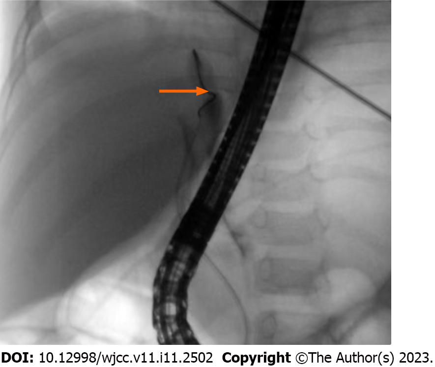
Figure 3 Dilated bile duct in the porta hepatis with significant distal stenosis (arrow), as observed via pressurized contrast imaging.
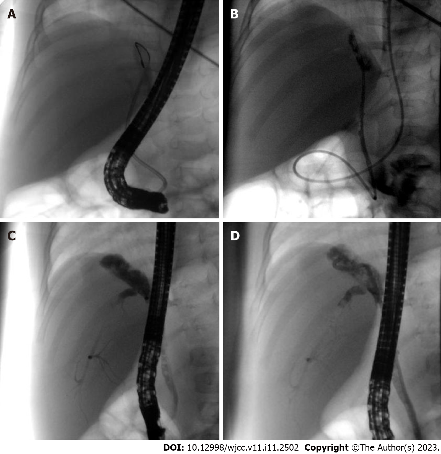
Figure 4 Treatment of the patient.
A: Inability and difficulty in passing the stenosis using a 7 Fr nasobiliary drainage tube; B: Smooth passage of a 5 Fr nasopancreatic tube through the stenosis; C: Dilatation of the bile ducts in the porta hepatis with visualization of the intrahepatic bile duct and the middle and lower common bile ducts; D: Smooth placement of a 7 Fr nasobiliary drainage tube.
- Citation: Shu J, Yang H, Yang J, Bian HQ, Wang X. Application of endoscopic retrograde cholangiopancreatography for treatment of obstructive jaundice after hepatoblastoma surgery: A case report. World J Clin Cases 2023; 11(11): 2502-2509
- URL: https://www.wjgnet.com/2307-8960/full/v11/i11/2502.htm
- DOI: https://dx.doi.org/10.12998/wjcc.v11.i11.2502


