Copyright
©The Author(s) 2023.
World J Clin Cases. Apr 6, 2023; 11(10): 2267-2275
Published online Apr 6, 2023. doi: 10.12998/wjcc.v11.i10.2267
Published online Apr 6, 2023. doi: 10.12998/wjcc.v11.i10.2267
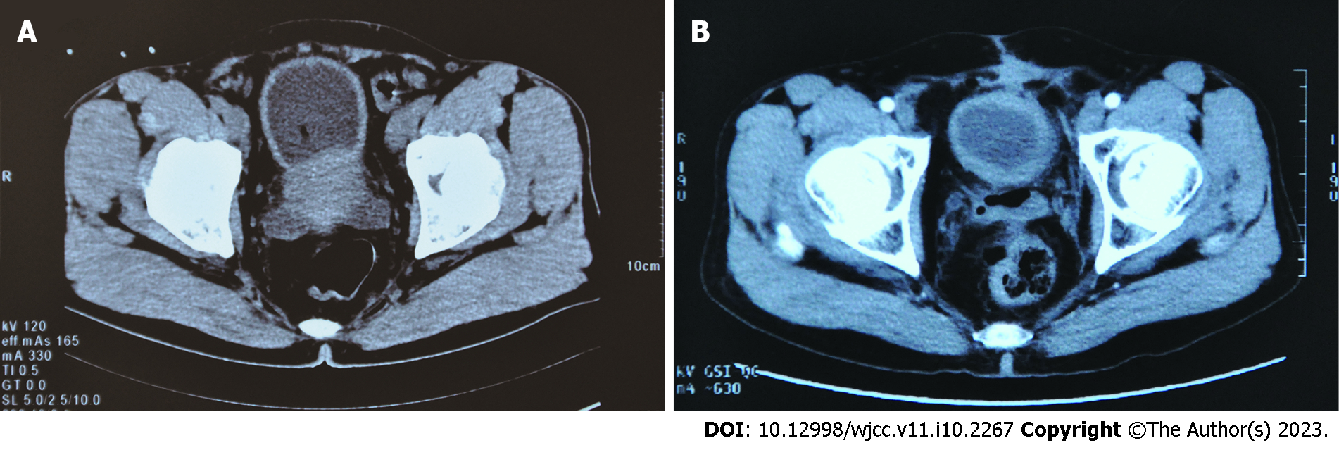
Figure 1 Computed tomography images of the patient before and after treatment.
A: Pelvic computed tomography (CT) image reveals an irregularly heterogeneity enlarged prostate with a rough surface involving the bladder neck and thickening of the bladder wall; B: Pelvic CT image shows no residue or recurrence of the tumor (10 mo after postoperative radiotherapy).
Figure 2 Histological image showing diffuse distribution of plasmacytoid/clear cytoplasm tumor cells with round to polygonal nuclei with prominent nucleoli; note admixed lymphocytes (hematoxylin-eosin staining, scale bar represents 200 μm).
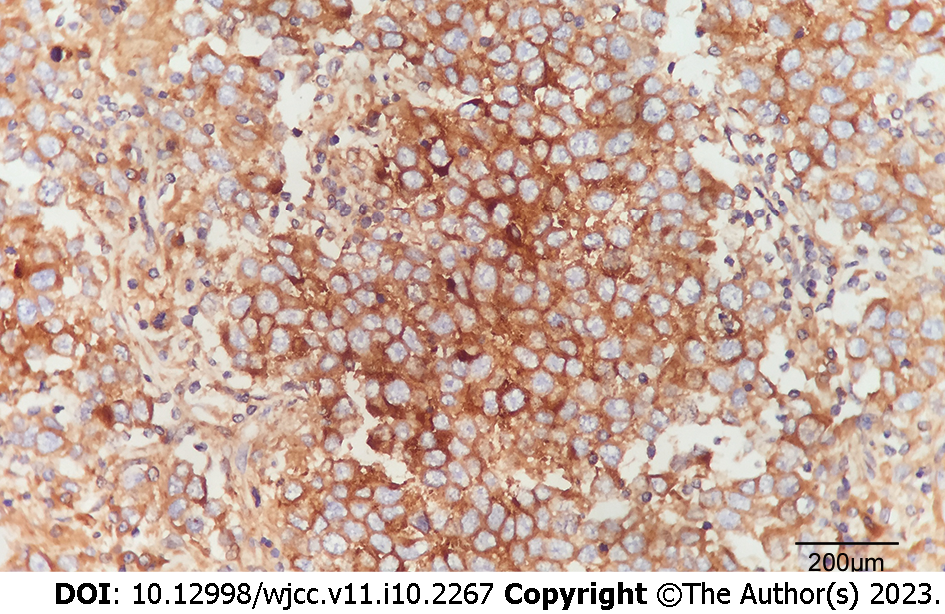
Figure 3 Immunohistochemical image showing strong membranous and cytoplasmic reactiveity for placental alkaline phosphatase in seminoma tumor cells (scale bar represents 200 μm).
- Citation: Cao ZL, Lian BJ, Chen WY, Fang XD, Jin HY, Zhang K, Qi XP. Diagnosis and treatment of primary seminoma of the prostate: A case report and review of literature. World J Clin Cases 2023; 11(10): 2267-2275
- URL: https://www.wjgnet.com/2307-8960/full/v11/i10/2267.htm
- DOI: https://dx.doi.org/10.12998/wjcc.v11.i10.2267


