Copyright
©The Author(s) 2022.
World J Clin Cases. Mar 16, 2022; 10(8): 2604-2609
Published online Mar 16, 2022. doi: 10.12998/wjcc.v10.i8.2604
Published online Mar 16, 2022. doi: 10.12998/wjcc.v10.i8.2604
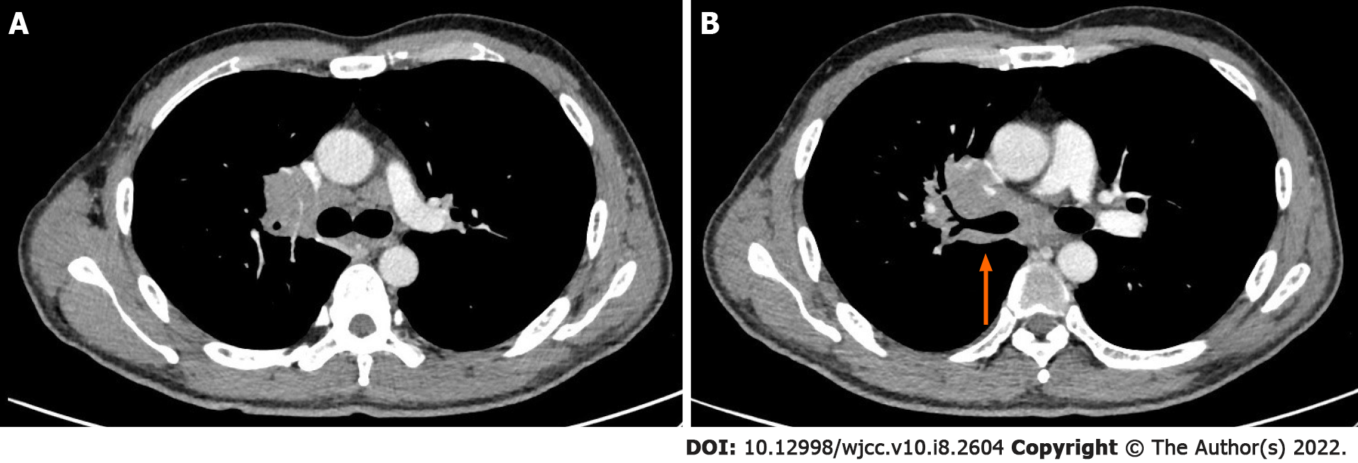
Figure 1 Chest enhanced computed tomography scan.
A: Right central type lung mass with enlarged lymph nodes in the right lung hilar, mediastinal and bilateral axillary areas; B: The computed tomography scan also displayed thickening of the right bronchial wall.
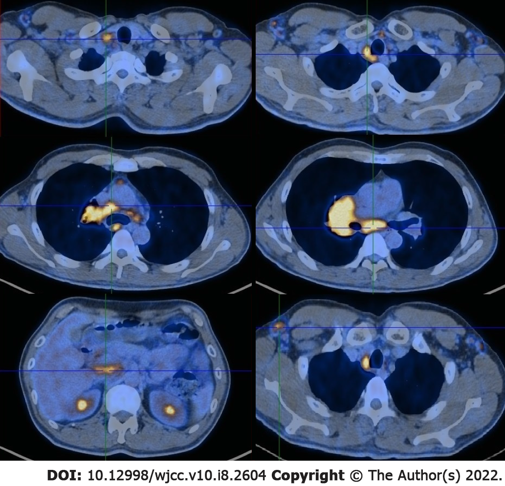
Figure 2
Positron emission tomography - computed tomography showed an increased uptake of 18-fludeoxyglucose in the mass of the right lung hilum and revealed lymph node metastases in the right lung hilar, mediastinal, celiac, right cervical, right supraclavicular and bilateral axillary areas.
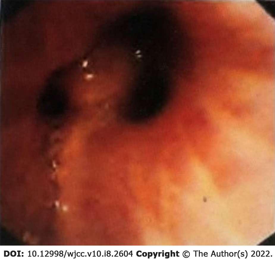
Figure 3
Flexible bronchoscopy displayed mucosal infiltrative changes at the level of the right upper lobe inlet and the right upper lobe bronchus was obstructed.
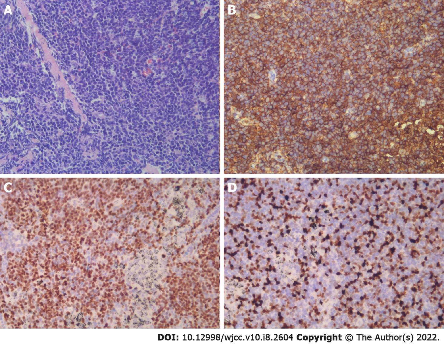
Figure 4 Mucosal endobronchial biopsy at the level of the right upper lobe inlet showing lymphoid hyperplasia.
A: H&E staining; B and C: With immunohistochemical staining, these cells were found to be positive for CD5 and SOX11; D: The percentage of Ki67-positive cells was 25% (Original magnification × 200).
- Citation: Ding YZ, Tang DQ, Zhao XJ. Mantle cell lymphoma with endobronchial involvement: A case report. World J Clin Cases 2022; 10(8): 2604-2609
- URL: https://www.wjgnet.com/2307-8960/full/v10/i8/2604.htm
- DOI: https://dx.doi.org/10.12998/wjcc.v10.i8.2604


