Copyright
©The Author(s) 2022.
World J Clin Cases. Feb 16, 2022; 10(5): 1738-1746
Published online Feb 16, 2022. doi: 10.12998/wjcc.v10.i5.1738
Published online Feb 16, 2022. doi: 10.12998/wjcc.v10.i5.1738
Figure 1 Macropathological and histological analyses of the tumour tissue in case 1.
A: Macroscopic image of the verrucous bulge; B-D: Haematoxylin and eosin staining showing the tumour cells (× 200); E-H: Positive immunohistochemical staining for CD10, CD99, transcription factor binding to IGHM enhancer-3 and CD163 (× 200); I-N: Negative immunohistochemical staining for S-100, cytokeratin, EMA, smooth muscle actin, Desmin, Stat6, ALK and NSE (× 200); O and P: Immunohistochemical staining for CD34 and Ki-67 (× 200).
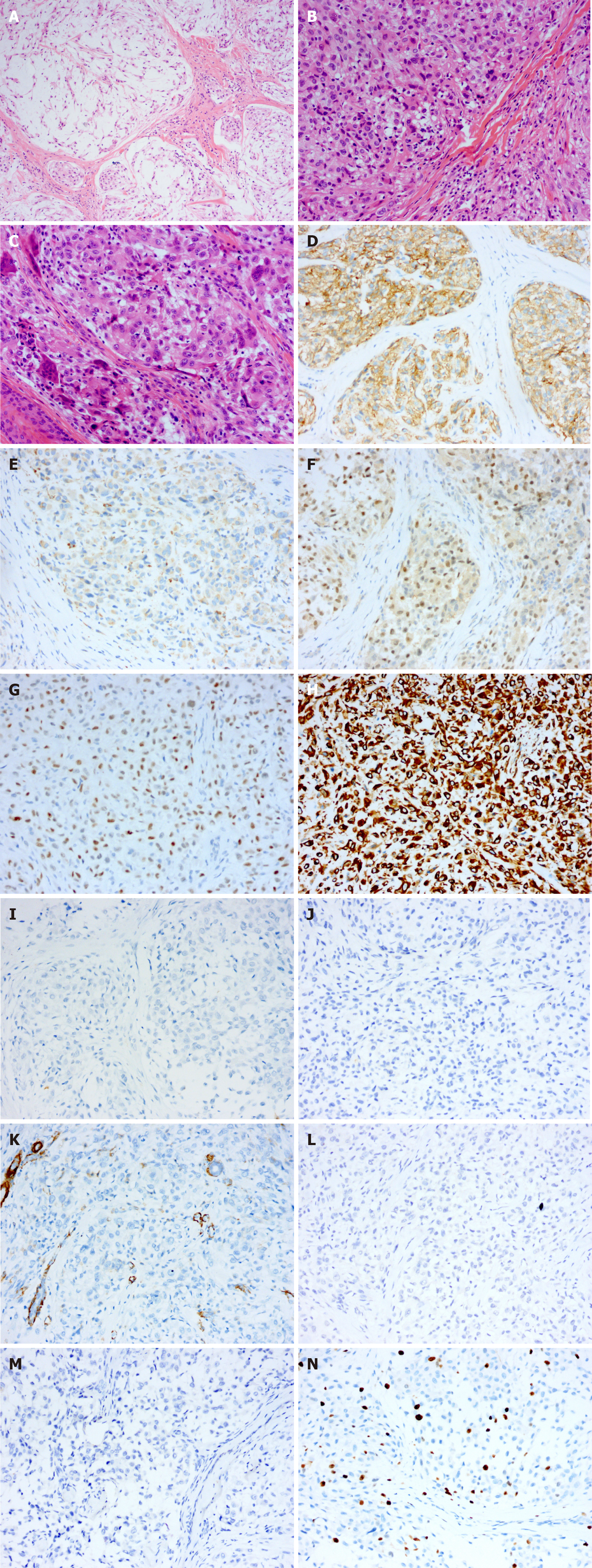
Figure 2 Histological analysis of the tumour tissue in case 2.
A-C: Haematoxylin and eosin staining showing the tumour cells (× 200); D-H: Positive immunohistochemical staining for CD10, CD68, transcription factor binding to IGHM enhancer-3, p63 and vimentin (× 200); I-M: Negative immunohistochemical staining for S-100, cytokeratin, smooth muscle actin, glial fibrillary acidic protein and CD1a (× 200); N: Immunohistochemical staining for Ki-67 (× 200).
- Citation: Huang WY, Zhang YQ, Yang XH. Neurothekeoma located in the hallux and axilla: Two case reports. World J Clin Cases 2022; 10(5): 1738-1746
- URL: https://www.wjgnet.com/2307-8960/full/v10/i5/1738.htm
- DOI: https://dx.doi.org/10.12998/wjcc.v10.i5.1738


