Copyright
©The Author(s) 2022.
World J Clin Cases. Feb 16, 2022; 10(5): 1623-1629
Published online Feb 16, 2022. doi: 10.12998/wjcc.v10.i5.1623
Published online Feb 16, 2022. doi: 10.12998/wjcc.v10.i5.1623
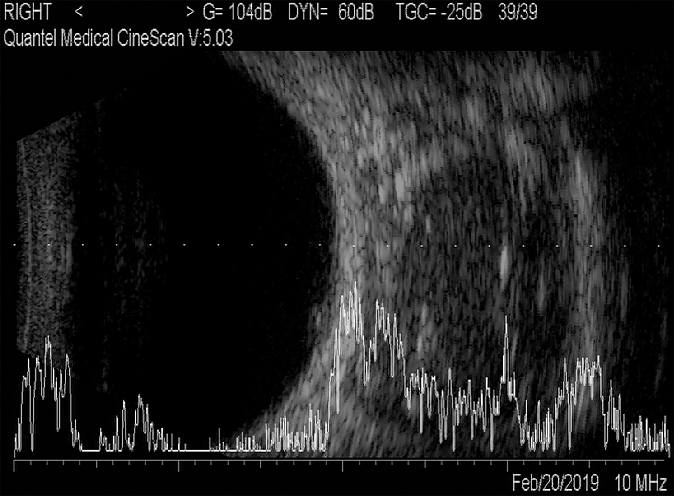
Figure 1 A/B-scan showed moderate echogenic lesions in the right eye orbital.
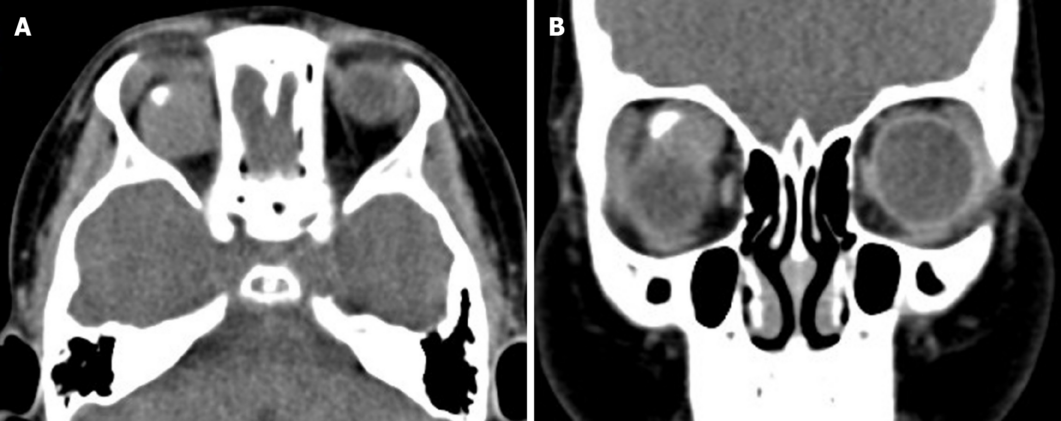
Figure 2 Orbital computed tomography scan showed a well-defined soft tissue density mass in the right orbit, with a hyperdense speck suggestive of coarse calcification.
A: Axial computed tomography (CT) scans; B: Coronal CT scans.
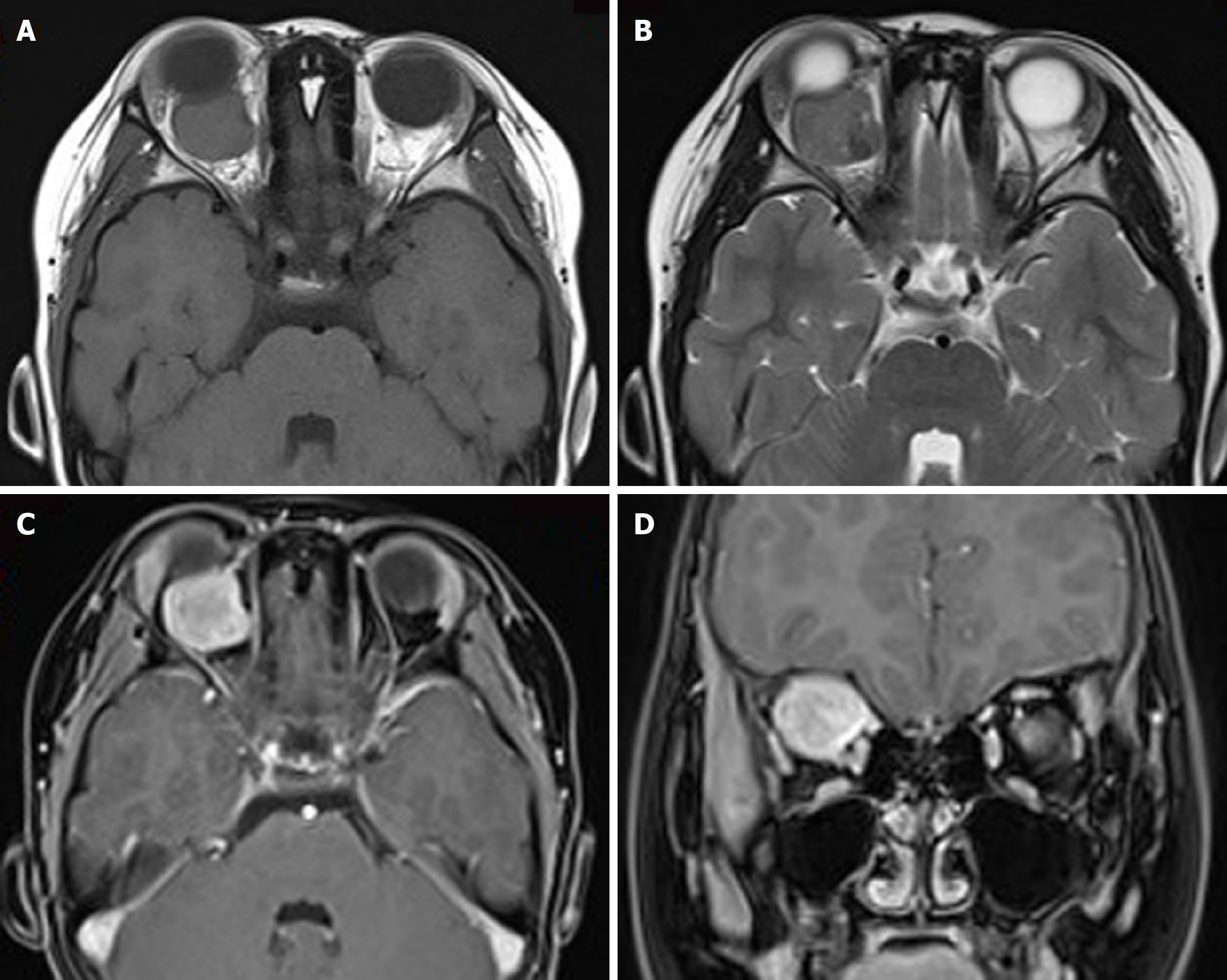
Figure 3 Orbital magnetic resonance imaging showed a circular-like mass in the right orbital.
A: T1-weighted images showed moderate signals, mixed with low-signal regions; B: T2-weighted images showed mixed signals, with high number of moderately high signals, and mixed with low-signal regions; C and D: Most part of the lesion was significantly and unevenly enhanced, whereas local lesions did not exhibit any enhancement.
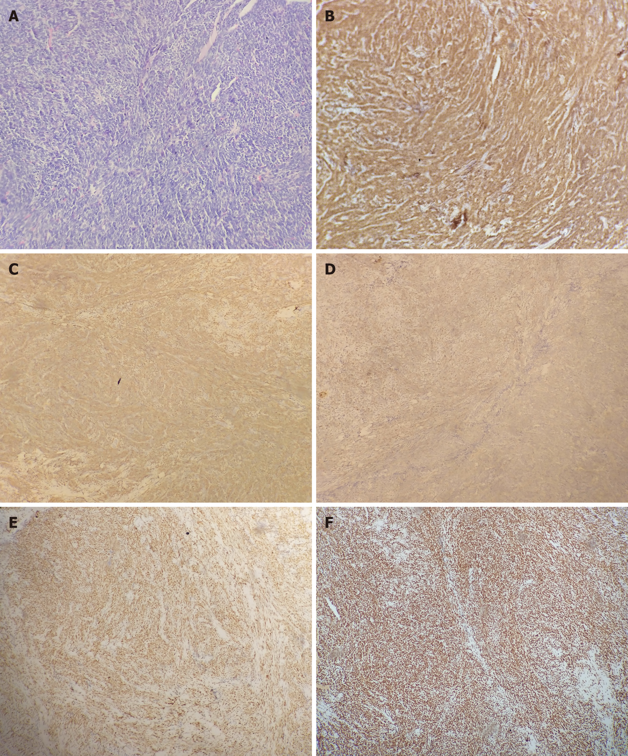
Figure 4 Histological and immunohistochemical examination.
A: The tumor was confirmed as monophasic synovial sarcoma with calcification based on histological analysis (HE, × 200); B-F: Immunohistochemical study revealed positive staining for Bcl-2 (B), CD99 (C), CKpan (D), TLE1 (E), and INI-1 (F) (× 100).
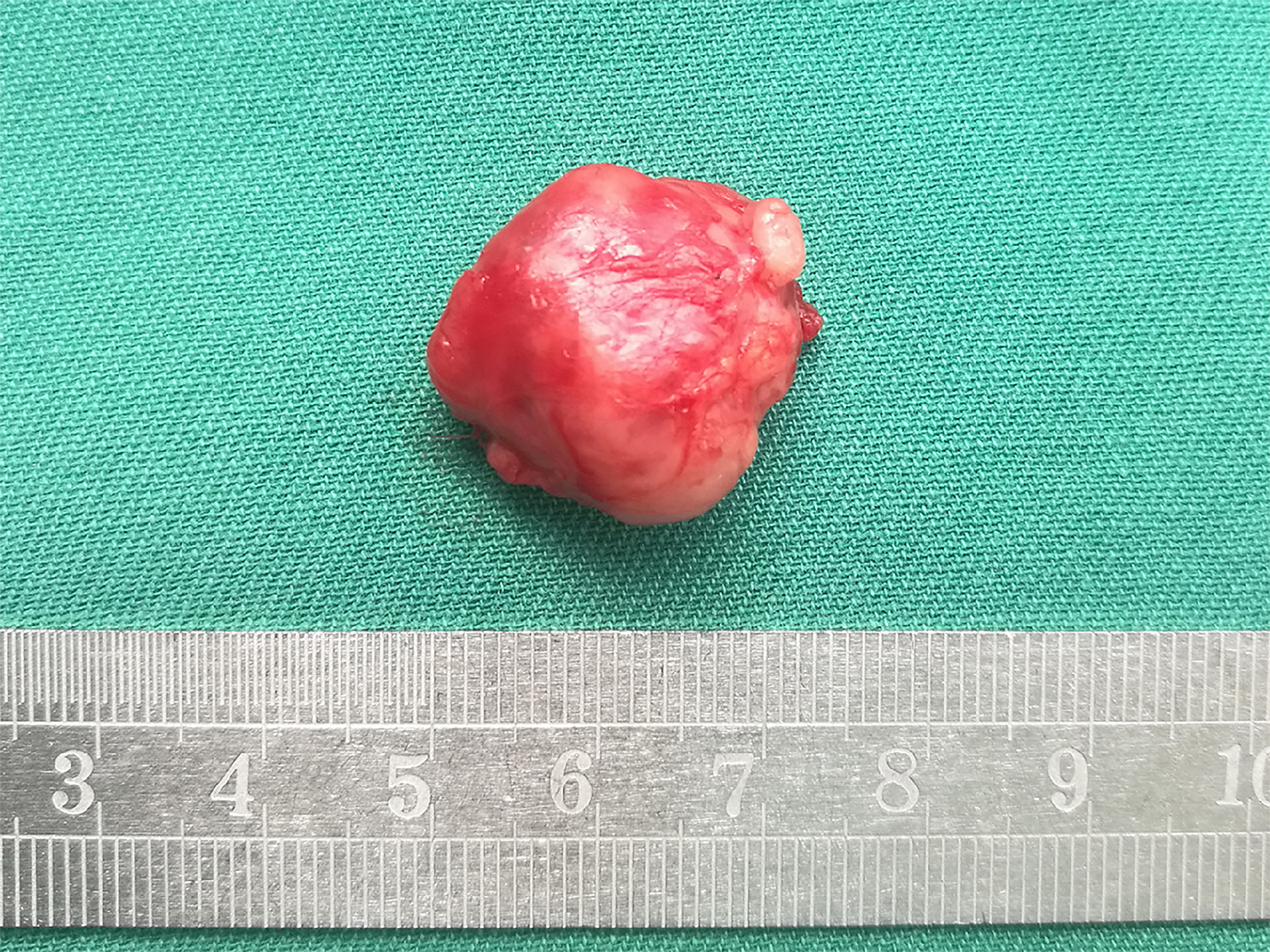
Figure 5 It presented as a well-defined, reddish, with irregularly shaped soft tissue density mass.
- Citation: Ren MY, Li J, Li RM, Wu YX, Han RJ, Zhang C. Primary orbital monophasic synovial sarcoma with calcification: A case report. World J Clin Cases 2022; 10(5): 1623-1629
- URL: https://www.wjgnet.com/2307-8960/full/v10/i5/1623.htm
- DOI: https://dx.doi.org/10.12998/wjcc.v10.i5.1623


