Copyright
©The Author(s) 2022.
World J Clin Cases. Feb 16, 2022; 10(5): 1592-1597
Published online Feb 16, 2022. doi: 10.12998/wjcc.v10.i5.1592
Published online Feb 16, 2022. doi: 10.12998/wjcc.v10.i5.1592
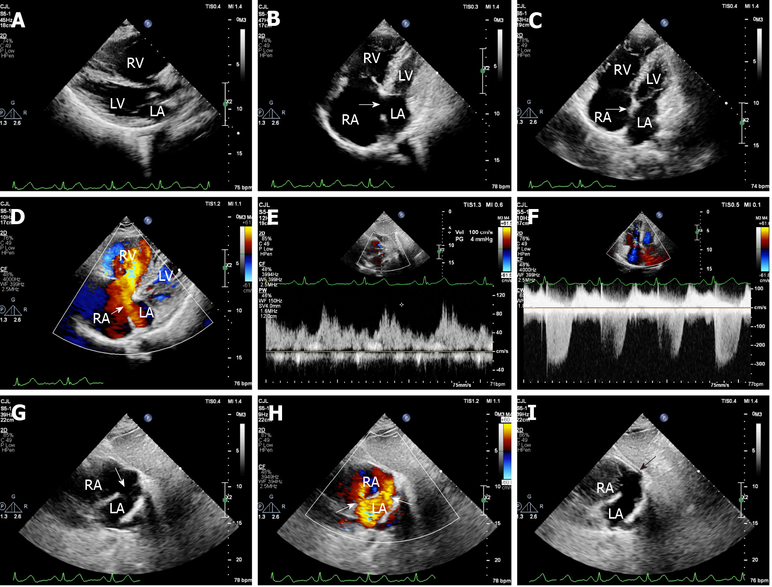
Figure 1 Transthoracic echocardiography before surgery.
A: Significant enlargement of the right ventricle (anterior-posterior diameter = 43 mm); B: Location of the defect was near the endocardial cushions on apical four-chamber view, which was mistaken for a defect of the ostium primum atrial septal defect (ASD) (arrow); C: When detected on apical four-chamber view by scanning backward, a defect of the coronary sinus (CS) in the terminal portion and normal endocardial cushions were seen (arrow); D: A shunt through the defect of the CS in the terminal portion on apical four-chamber view (arrow); E: Pulse-wave Doppler spectrum showed a shunt during diastole through the defect of the CS in the terminal portion [Vmax = 100 cm/s, pressure gradient (PG) = 4 mmHg]; F: Moderate-to-severe tricuspid regurgitation (Vmax = 337cm/s, PG = 45 mmHg, pulmonary artery systolic pressure = 50 mmHg); G: The defect of the CS in the terminal portion and secundum ASD on subxiphorid biatrial view (arrow, 3.3 cm × 2.0 cm and 1.1 cm); H: Two shunts through the defect of the CS and secundum ASD on subxiphorid biatrial view (arrow); I: Negative filling was observed at the right atrium orifice of the CS and right atrium side of the secundum atrial septal by right-heart contrast echocardiography (arrow). RA: Right atrium; RV: Right ventricle; LA: Left atrium; LV: Left ventricle.
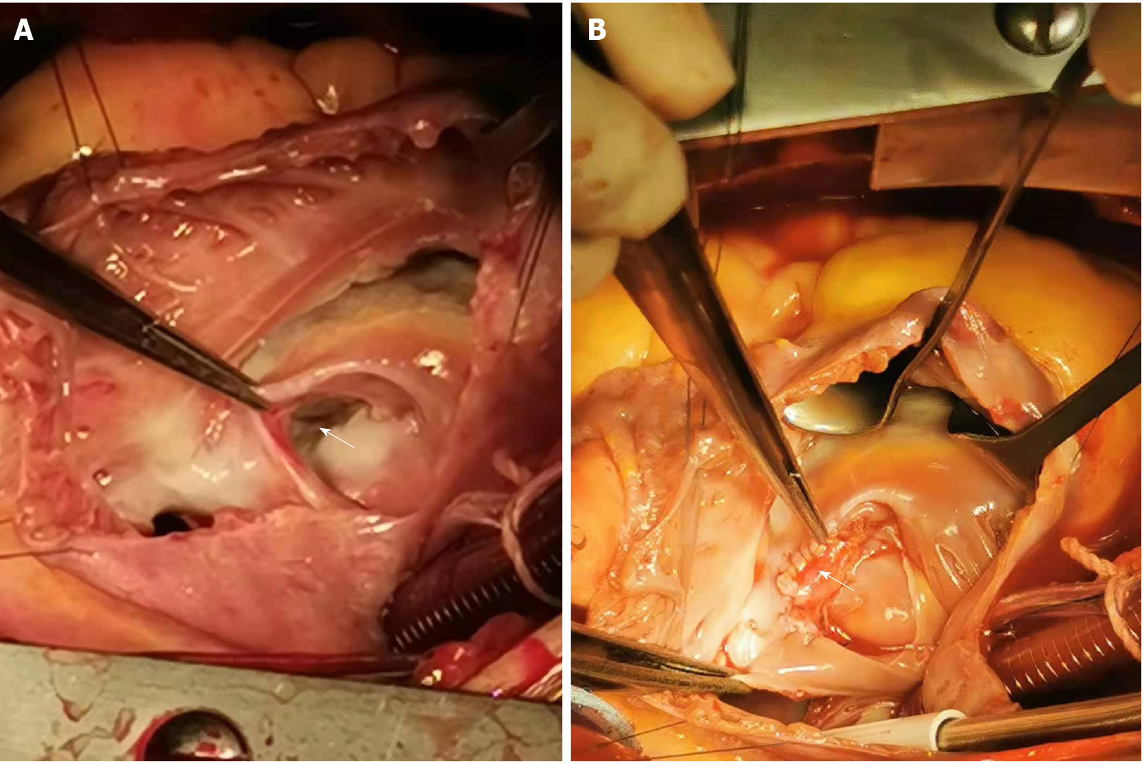
Figure 2 Imaging during the operation.
A: Obvious broadening of the coronary sinus (CS) with a partial defect of the CS roof in the terminal portion (3.0 cm × 2.1 cm) was seen upon incision of the right atrium; B: The defect of the CS in the terminal portion was repaired.
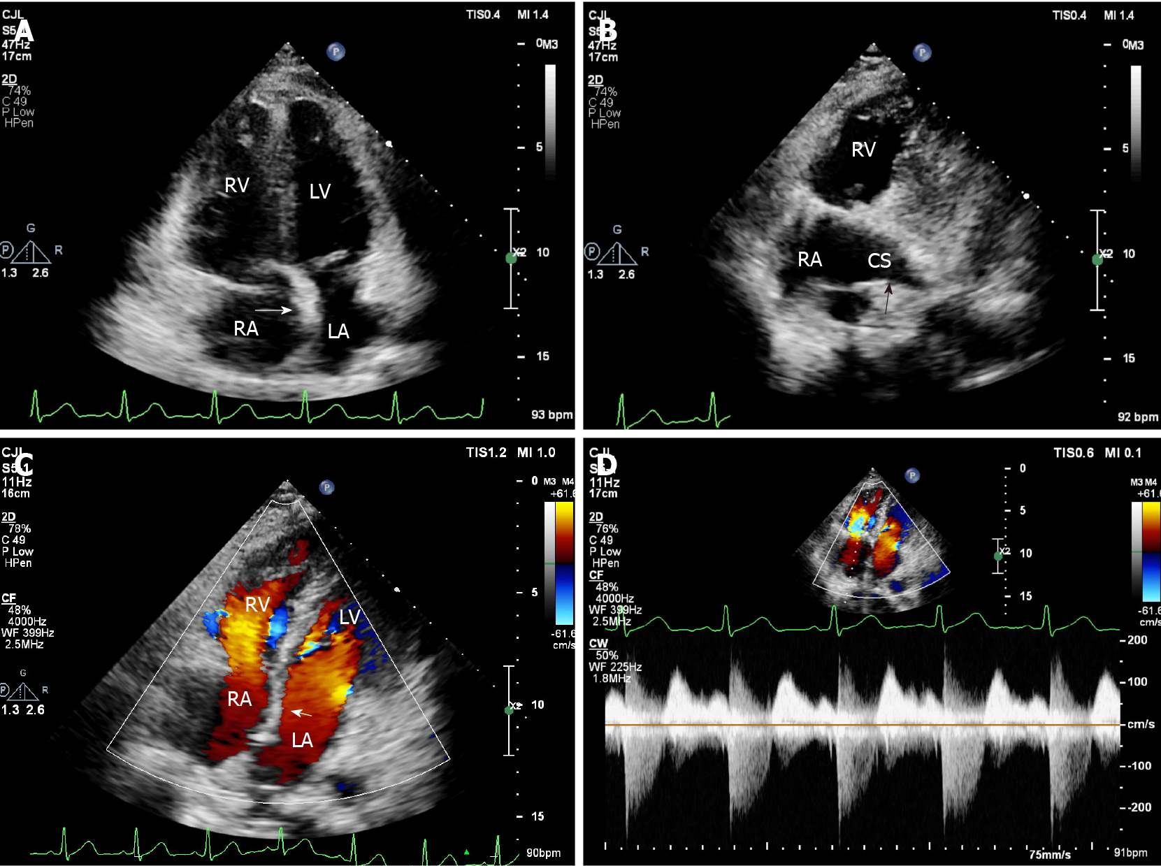
Figure 3 Transthoracic echocardiography at 1 wk after surgery.
A: The repaired atrial septum was continuous and complete on apical four-chamber view (arrow); B: The repaired coronary sinus (CS) roof was continuous and complete on apical four-chamber view (arrow); C: There was no shunt from the left atrium to right atrium on apical four-chamber view (arrow); D: Trace tricuspid regurgitation (Vmax = 223 cm/s, pressure gradient = 20 mmHg, pulmonary artery systolic pressure = 25 mmHg). RA: Right atrium; RV: Right ventricle; LA: Left atrium; LV: Left ventricle; CS: Coronary sinus.
- Citation: Chen JL, Yu CG, Wang DJ, Chen HB. Misdiagnosis of unroofed coronary sinus syndrome as an ostium primum atrial septal defect by echocardiography: A case report. World J Clin Cases 2022; 10(5): 1592-1597
- URL: https://www.wjgnet.com/2307-8960/full/v10/i5/1592.htm
- DOI: https://dx.doi.org/10.12998/wjcc.v10.i5.1592


