Copyright
©The Author(s) 2022.
World J Clin Cases. Dec 16, 2022; 10(35): 13081-13087
Published online Dec 16, 2022. doi: 10.12998/wjcc.v10.i35.13081
Published online Dec 16, 2022. doi: 10.12998/wjcc.v10.i35.13081
Figure 1 Slit lamp examination.
A: Anterior segment image of the affected eye; B: Koeppe nodules at the pupillary margin of the affected eye; C: Anterior segment image of the fellow eye.
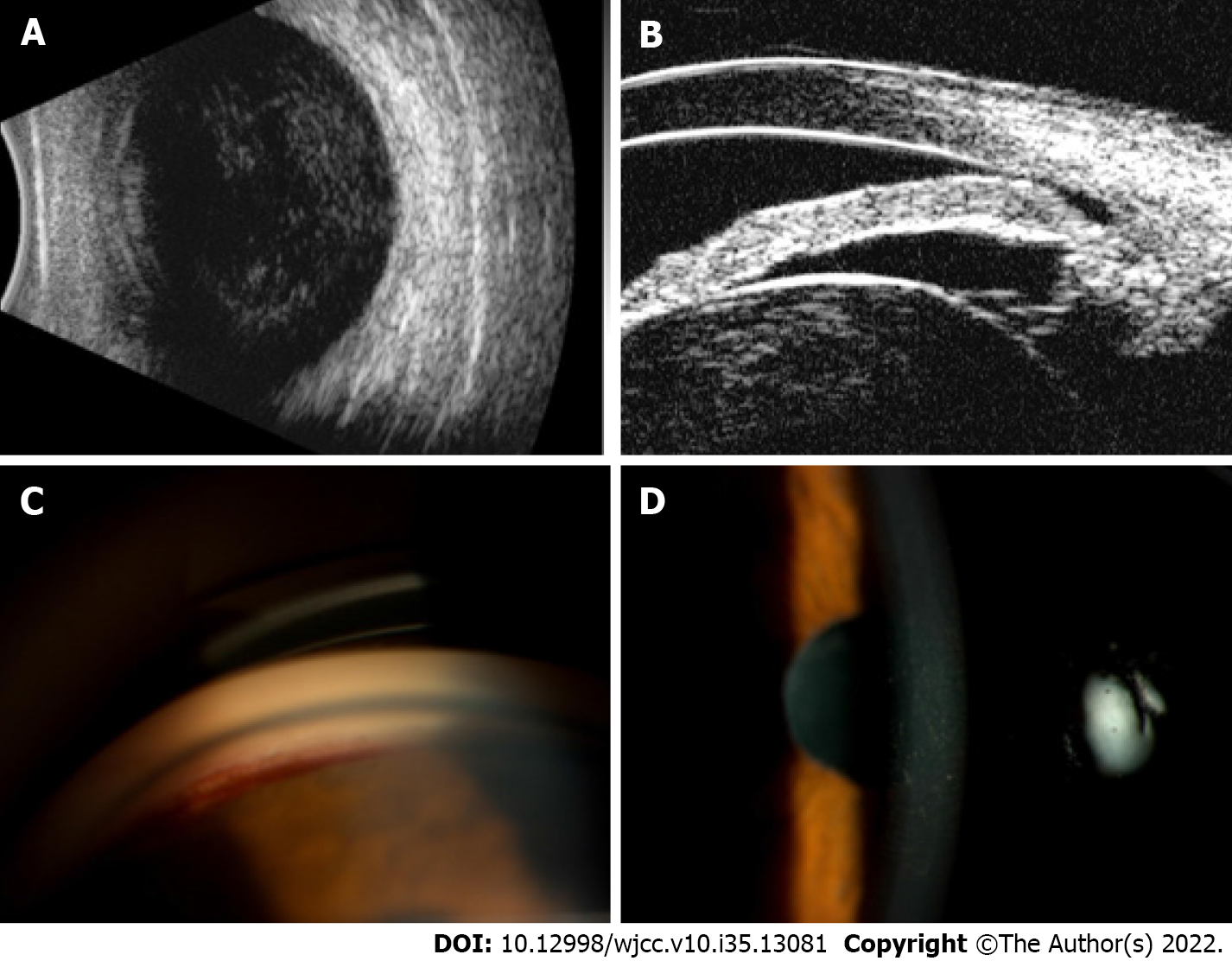
Figure 2 Clinical features of the affected eye.
A: B-ultrasound revealed vitreous opacity in the affected right eye; B: Ultrasound biomicroscopy showed a shallow anterior chamber and lens opacification and expansion; C: Fresh bleeding at the anterior chamber angle of the affected eye during gonioscopy; D: Hammered silver appearance of central corneal endothelium in the affected eye.
Figure 3 Specular microscopy examination.
A: Wide-band dark area of the affected eye revealed by specular microscopy; B: Normal endothelial morphology of the fellow eye.
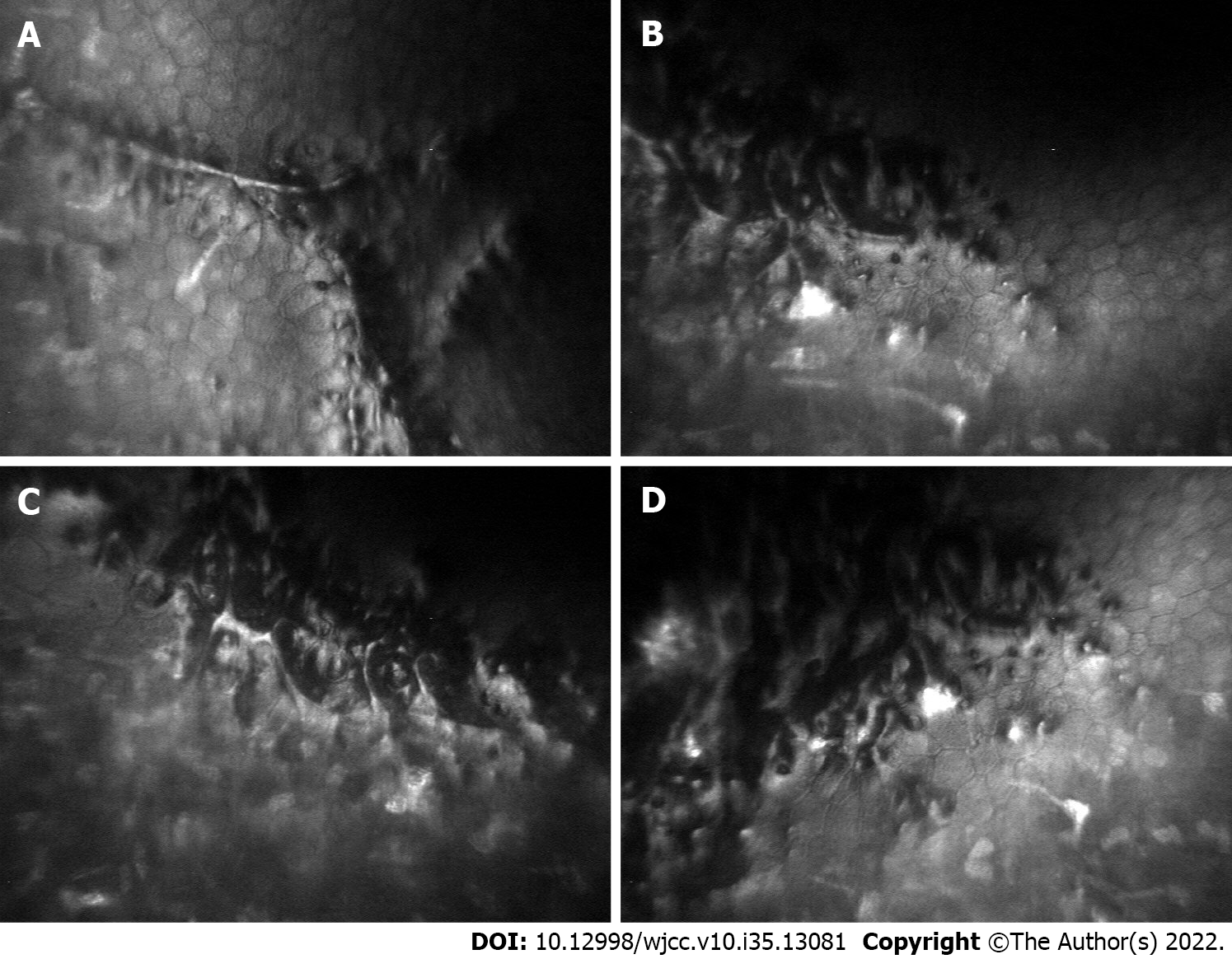
Figure 4 In vivo confocal microscopy examination.
A-D: Irregular corneal endothelial protuberances and dark bodies of various sizes in the affected eye (images from different areas) are visible.
- Citation: Cheng YY, Wang CY, Zheng YF, Ren MY. Hammered silver appearance of the corneal endothelium in Fuchs uveitis syndrome: A case report. World J Clin Cases 2022; 10(35): 13081-13087
- URL: https://www.wjgnet.com/2307-8960/full/v10/i35/13081.htm
- DOI: https://dx.doi.org/10.12998/wjcc.v10.i35.13081


