Copyright
©The Author(s) 2022.
World J Clin Cases. Dec 16, 2022; 10(35): 13028-13037
Published online Dec 16, 2022. doi: 10.12998/wjcc.v10.i35.13028
Published online Dec 16, 2022. doi: 10.12998/wjcc.v10.i35.13028
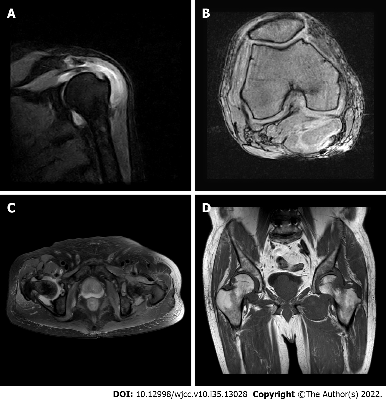
Figure 1 Case 1: Shoulder, hip, and knee magnetic resonance imaging in 2018.
A: Shoulder synovial hyperplasia; B: Knee meniscus injury; C and D: Femoral neck erosion with hyperplasia of the surrounding synovial tissue.
Figure 2 Case 2: Shoulder and wrist magnetic resonance imaging in 2017.
A and B: Acromion-deltoid subcapsular effusion; C and D: Wrist joint effusion with synovial tissue hyperplasia.
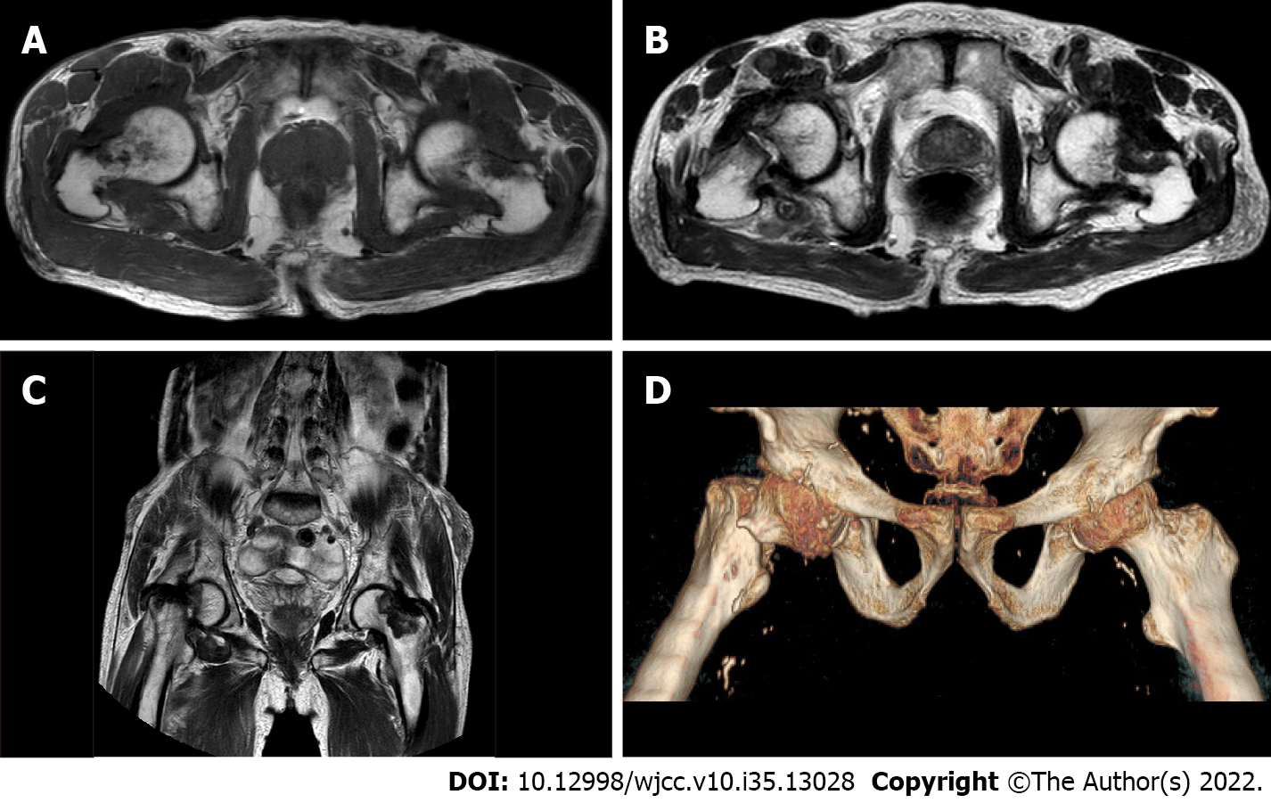
Figure 3 Case 2: Hip magnetic resonance imaging and 3D imaging.
A: Femoral neck erosion with surrounding synovial hyperplasia in 2018; B-D: Fracture of the right femoral neck in 2019.
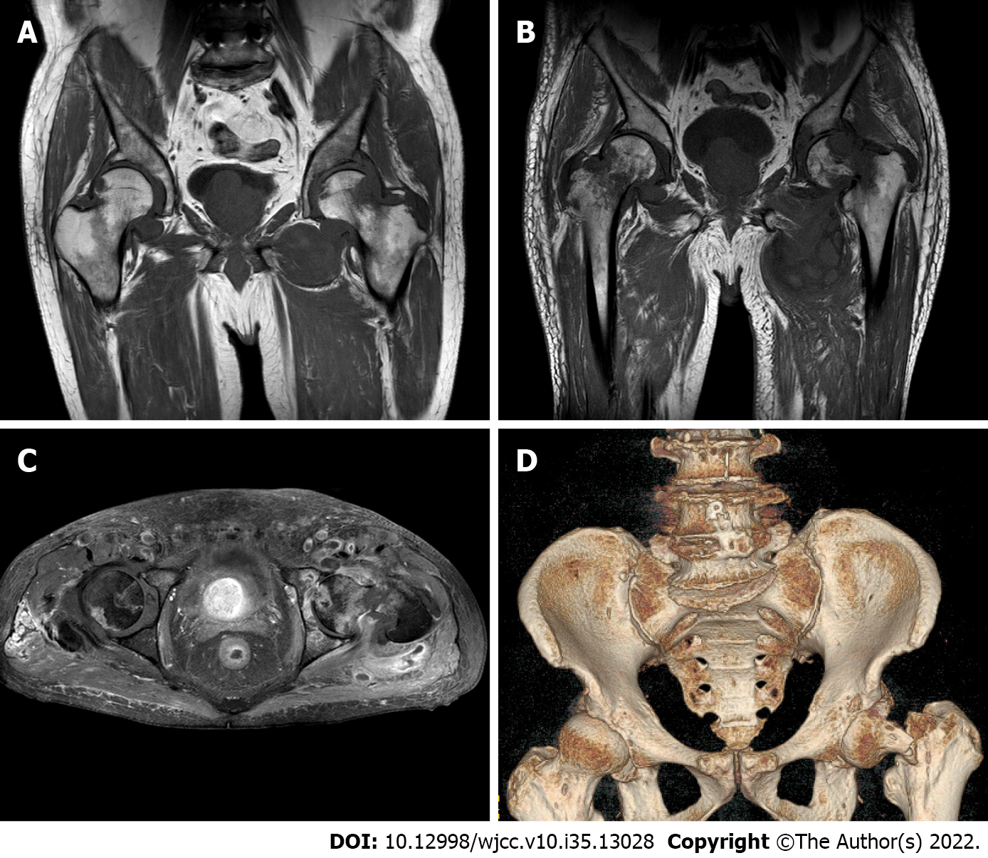
Figure 4 Case 1: Hip magnetic resonance imaging and 3D imaging in 2019.
A-C: Old fracture of the left femoral neck and synovial cyst around the left hip joint; D: Left femoral fracture.
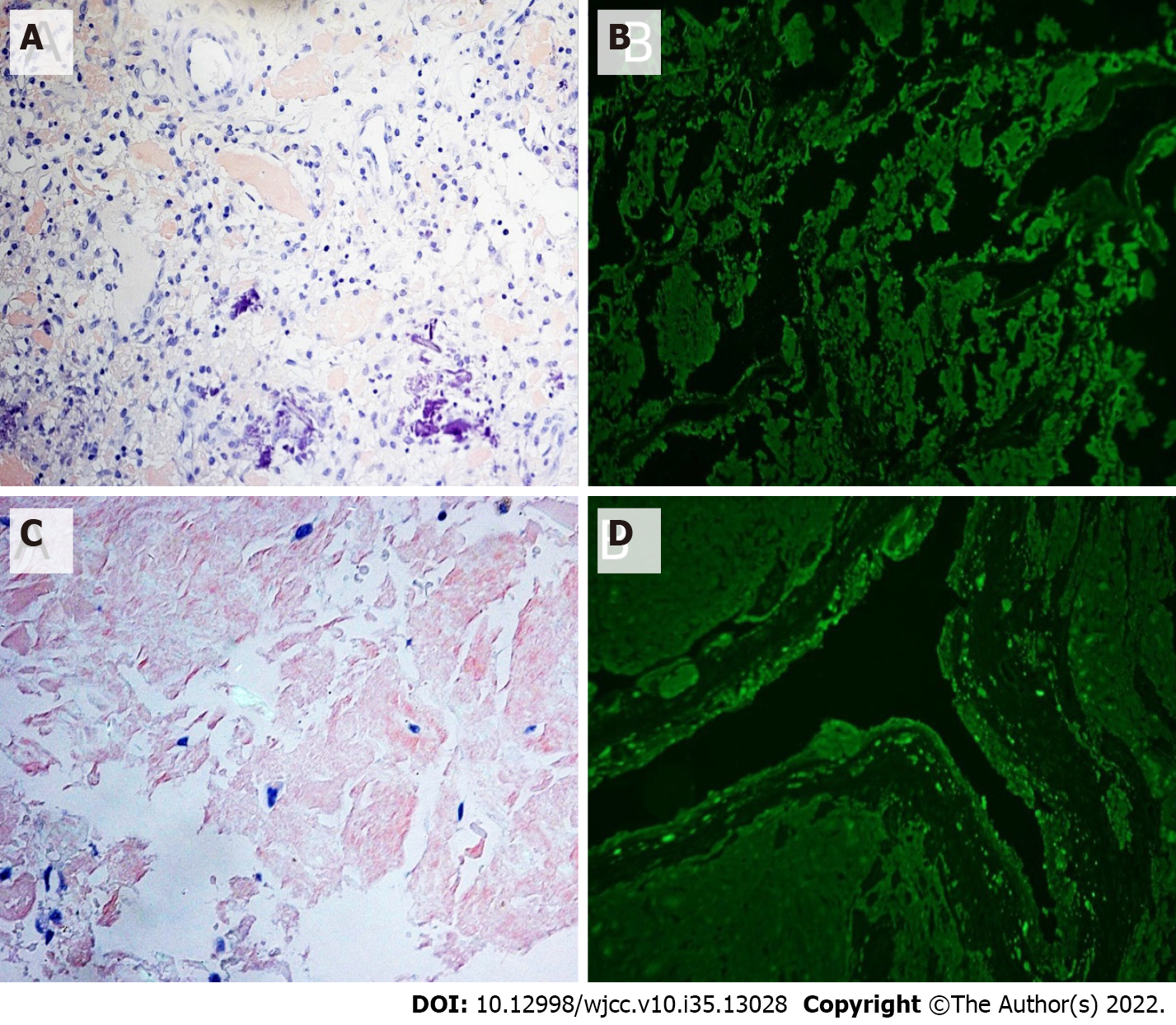
Figure 5 Case 1 and Case 2: Congo red staining and immunofluorescence.
A: Congo red staining ×200 (+); B: Immunofluorescence showing lambda light chain (+); C: Congo red staining ×200 (+); D: Immunofluorescence showing kappa light chain (+).
- Citation: He C, Ge XP, Zhang XH, Chen P, Li BZ. Multiple myeloma presenting with amyloid arthropathy as the first manifestation: Two case reports. World J Clin Cases 2022; 10(35): 13028-13037
- URL: https://www.wjgnet.com/2307-8960/full/v10/i35/13028.htm
- DOI: https://dx.doi.org/10.12998/wjcc.v10.i35.13028


