Copyright
©The Author(s) 2022.
World J Clin Cases. Nov 16, 2022; 10(32): 11869-11876
Published online Nov 16, 2022. doi: 10.12998/wjcc.v10.i32.11869
Published online Nov 16, 2022. doi: 10.12998/wjcc.v10.i32.11869
Figure 1 Light microscopy findings.
A: Many glomeruli had active global cellular crescents with segmental necrotizing lesions and polymorphonuclear leukocytes in the glomeruli. Hematoxylin and eosin (H&E), magnification 200 ×; B: Rare glomeruli that were not involved by crescents showed mesangial hypercellularity. H&E, magnification 200 ×.
Figure 2 Immunofluorescence studies.
A: No glomerular staining for albumin; B: Bright diffuse global linear staining for immunoglobulin (Ig)G was seen along the glomerular capillary loops; C: Moderate to prominent diffuse global granular staining for IgA was seen in the glomerular mesangium; D: Prominent diffuse global granular staining for C3 was seen in the glomerular mesangium.
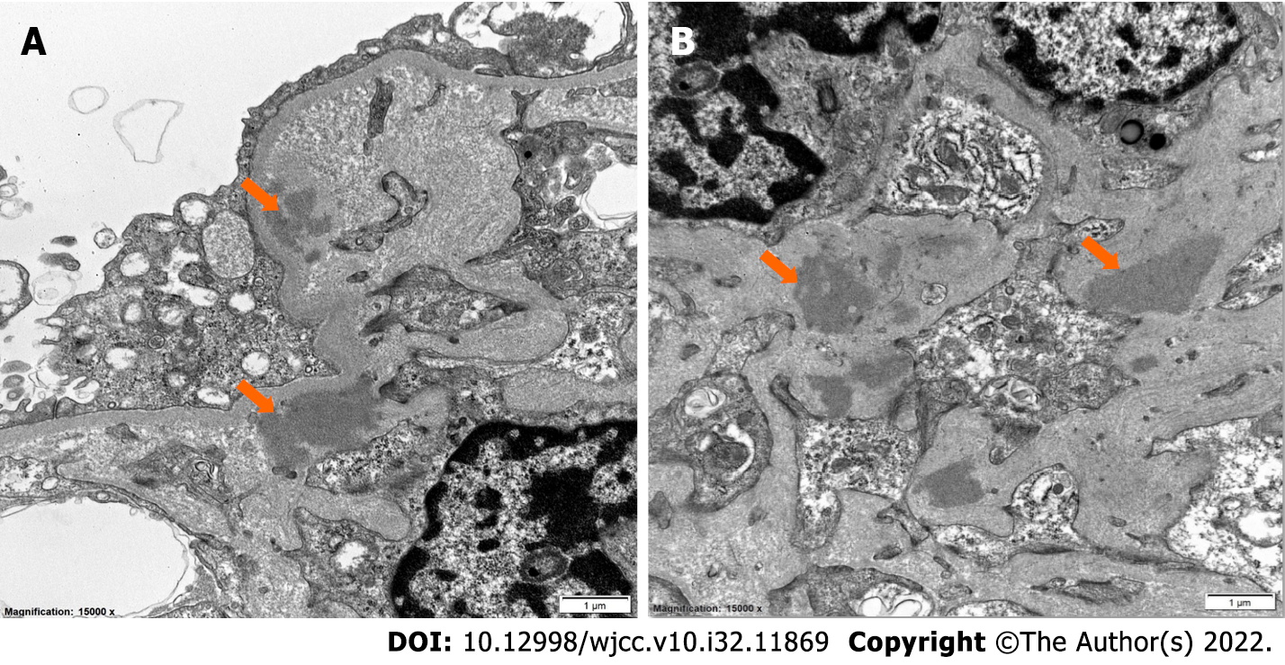
Figure 3 Ultrastructural examination.
A: Electron dense immune complex type deposits were seen in paramesangium (arrow); B: Electron dense immune complex type deposits were seen in the mesangium (arrow).
- Citation: Ibrahim D, Brodsky SV, Satoskar AA, Biederman L, Maroz N. Triple hit to the kidney-dual pathological crescentic glomerulonephritis and diffuse proliferative immune complex-mediated glomerulonephritis: A case report. World J Clin Cases 2022; 10(32): 11869-11876
- URL: https://www.wjgnet.com/2307-8960/full/v10/i32/11869.htm
- DOI: https://dx.doi.org/10.12998/wjcc.v10.i32.11869


