Copyright
©The Author(s) 2022.
World J Clin Cases. Oct 26, 2022; 10(30): 11146-11154
Published online Oct 26, 2022. doi: 10.12998/wjcc.v10.i30.11146
Published online Oct 26, 2022. doi: 10.12998/wjcc.v10.i30.11146
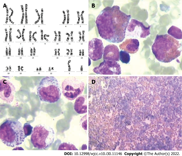
Figure 1 The patient’s karyotype is 46, XY, t (9;22) (q34;q11).
A: The typical translocation between the chromosomes 9 and 22 was found in all 20 metaphase cells analyzed; B-D: Bone marrow smear shows an extremely active cellular proliferation, myeloid cells that are dominated by myelocytes and metamyelocytes, and easily observable/high rate of eosinophils and basophils.
Figure 2 Computed tomography images show gastric cancer.
A: Computed tomography (CT) in venous phase show edematous and thickened gastric wall; B: CT in the arterial phase, the gastric wall was thickened and significantly enhanced.
Figure 3
Emission computed tomography images show abnormal bone metabolism in the bilateral scapulae, ribs, and ilium; multiple bone metastases were considered.
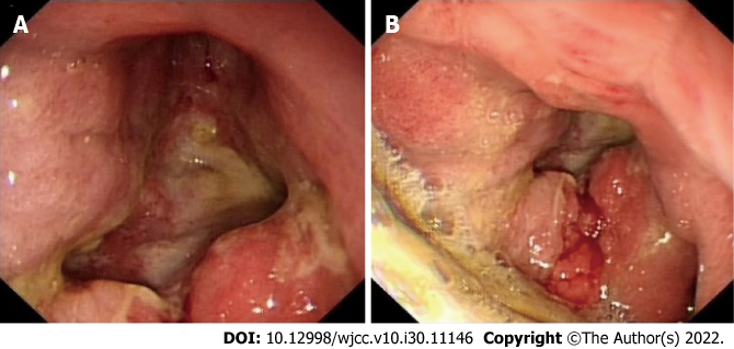
Figure 4 Gastroscopic images show gastric cancer (Bowman type IV).
A: A large amount of retained material in the gastric cavity and huge ulcers in the lower part of the gastric body, angle, and antrum; B: The ulcers base is uneven, brittle, and easy to bleed. Local peristalsis has disappeared.
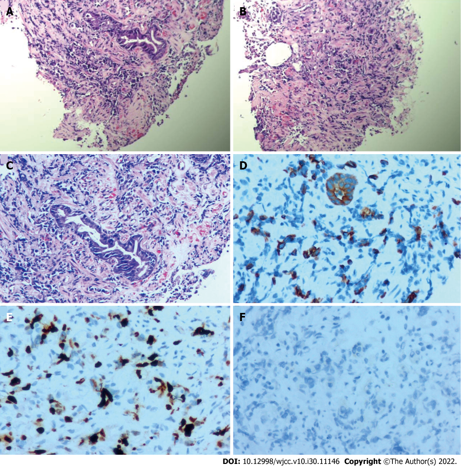
Figure 5 Pathological examination after gastroscopic biopsy reveals the presence of a small number of atypical cells.
A-C: Hematoxylin-eosin staining of biopsy specimens indicate poorly differentiated adenocarcinoma (A, 40 ×; B, 40 ×; C, 100 ×); D-F: Immunohistochemical staining shows that the specimens are positive for CKL (D, 200 ×), and Ki-65 (E, 200 ×) and negative for Her-2 (F, 200 ×).
Figure 6 Computed tomography images.
A: Computed tomography images show significantly thickened gastric wall; B: The tumor has invaded the hepatic flexure of the transverse colon, resulting in marked dilation of the upper bowel.
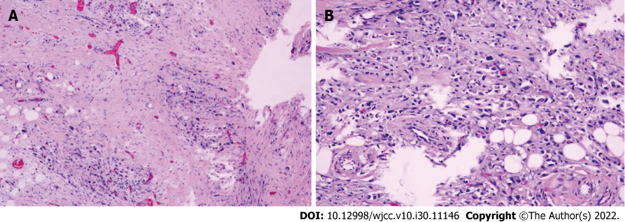
Figure 7 Histopathological examination shows a small mesenteric nodule, likely metastatic adenocarcinoma.
A: Hematoxylin-eosin stain: 40 ×; B: Hematoxylin-eosin stain: 100 ×.
- Citation: Zhao YX, Yang Z, Ma LB, Dang JY, Wang HY. Synchronous gastric cancer complicated with chronic myeloid leukemia (multiple primary cancers): A case report. World J Clin Cases 2022; 10(30): 11146-11154
- URL: https://www.wjgnet.com/2307-8960/full/v10/i30/11146.htm
- DOI: https://dx.doi.org/10.12998/wjcc.v10.i30.11146


