Copyright
©The Author(s) 2022.
World J Clin Cases. Oct 16, 2022; 10(29): 10742-10754
Published online Oct 16, 2022. doi: 10.12998/wjcc.v10.i29.10742
Published online Oct 16, 2022. doi: 10.12998/wjcc.v10.i29.10742
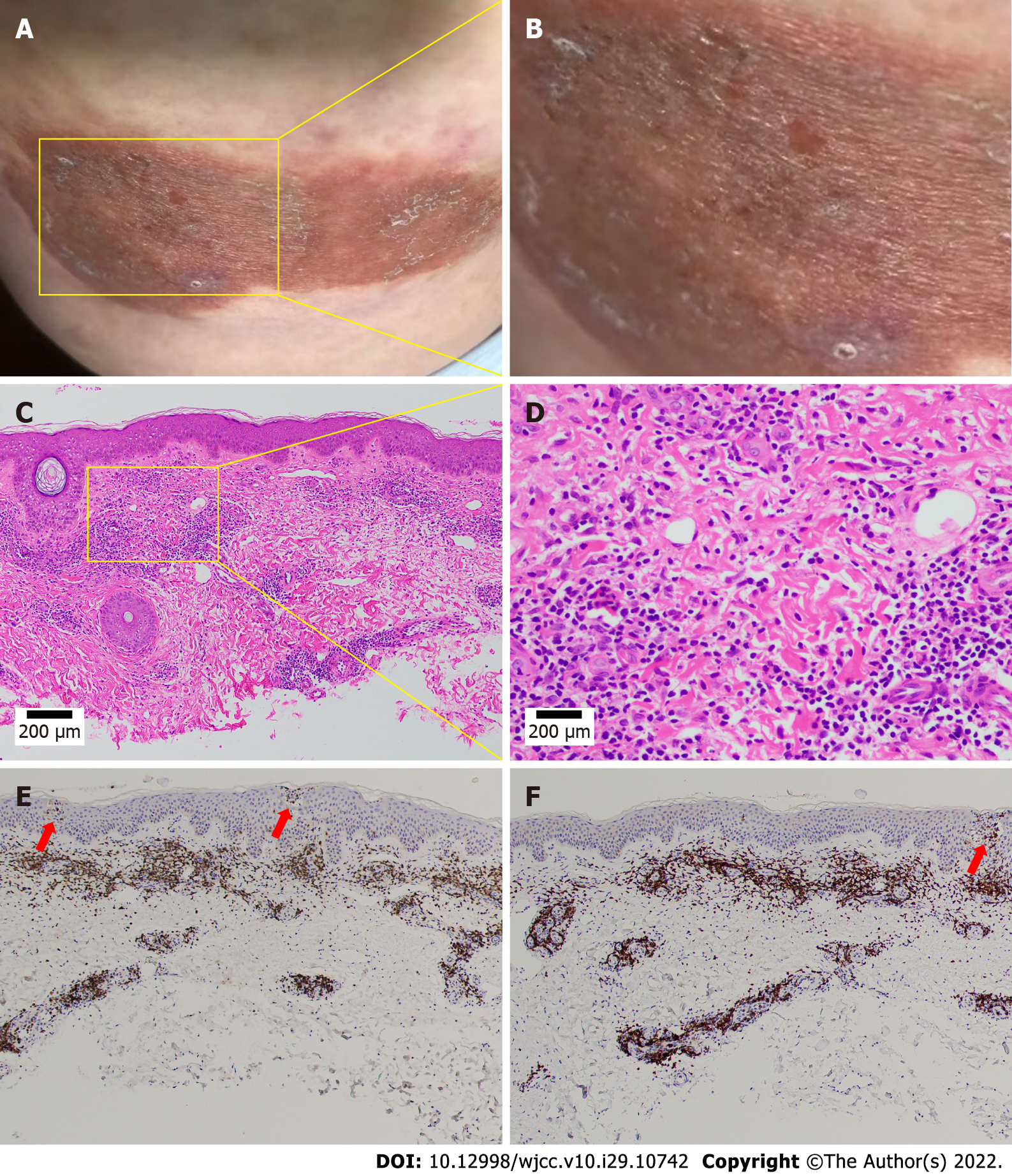
Figure 1 Clinical presentation and diagnostic clues from the original symptoms.
A and B: Intensely pruritic, weeping, erythematous, eczematous, and indurated plaques; C: An intense perivascular infiltrate composed of lymphocytes and histiocytic cells, with rare plasma cells and eosinophils (hematoxylin–eosin stain, 100 ×); D: Prominent endothelial cell swelling. The infiltrate extended through the reticular dermis into the subcutaneous fat and focally surrounded the eccrine and neural structures (hematoxylin–eosin stain, 400 ×); E and F: The lower half of the dermis was markedly sclerotic with thickened eosinophilic collagen bundles. A microscopic examination revealed a normal epidermis and more lymphocytes infiltrating the blood vessels in the dermis, several of which were mimicking Pautrier microabscesses (100 ×).
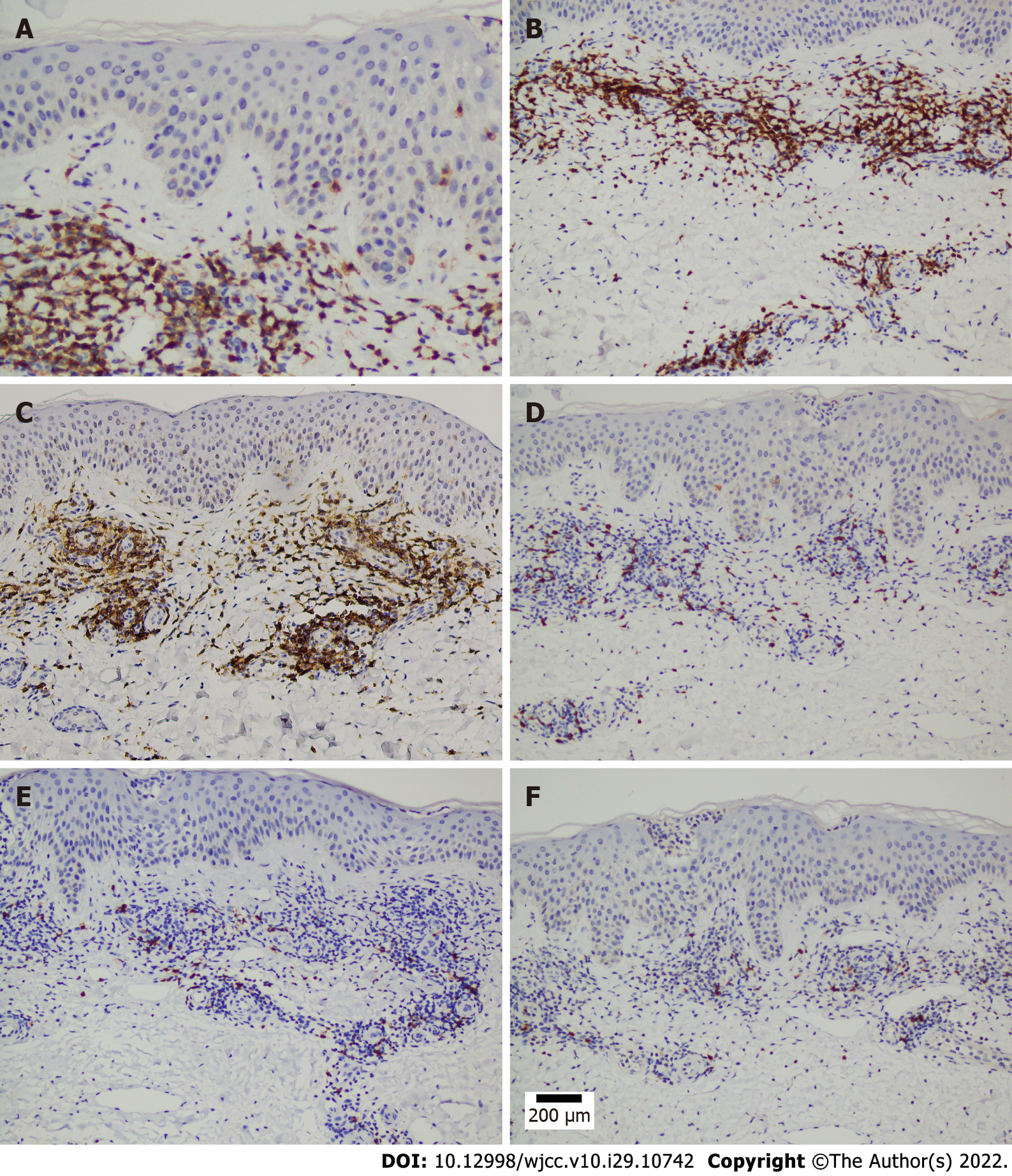
Figure 2 Immunohistochemical assay for positive phenotypes.
A-D: T cells tested positive for CD3, CD5, and CD4 and partially positive for CD8 (200 ×); E and F: B cells tested positive for CD20 and CD79a (200 ×).
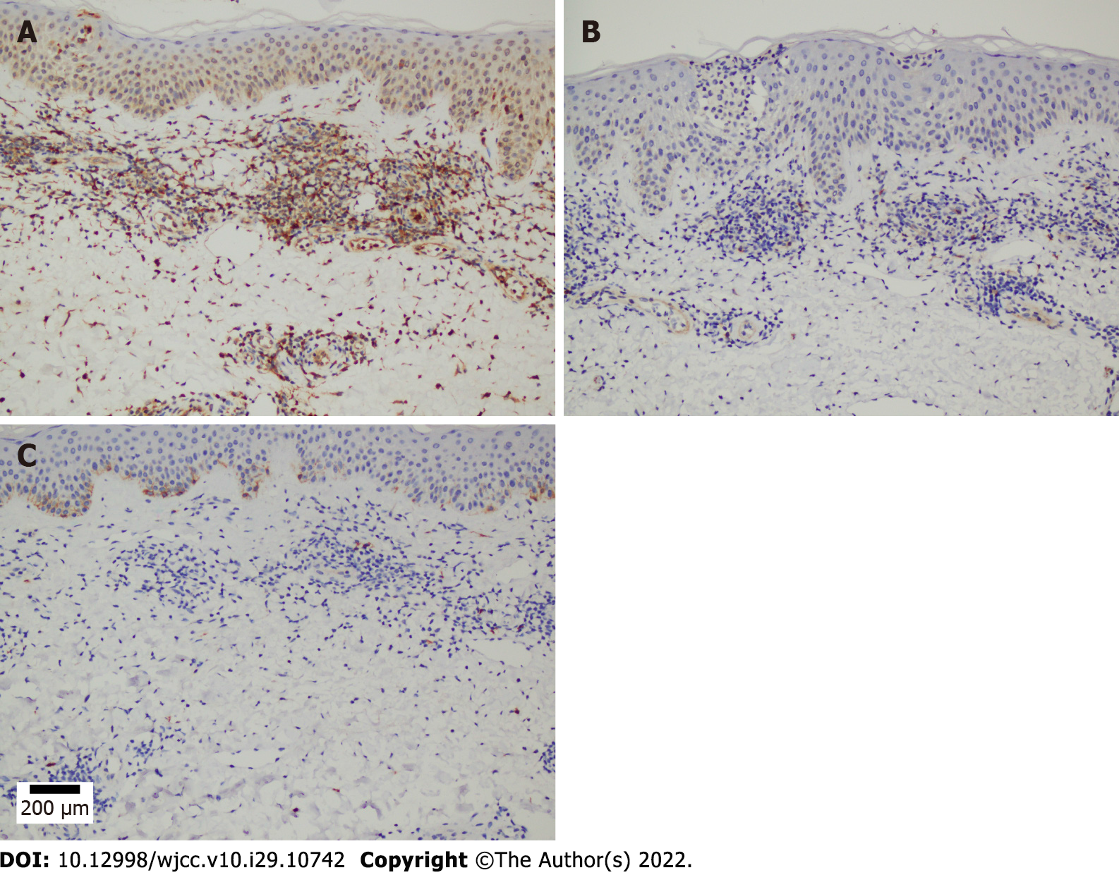
Figure 3 Immunohistochemical assays for other phenotypes.
A-C: Histocytes were positive for CD68+, natural killer cells tested positive for CD56, and mastocytes tested negative for CD117 (200 ×).
Figure 4 Clinical presentation before and after treatment.
A: Appearance of the skin before treatment (September 14, 2021); B: Appearance of the skin during follow-up (March 6, 2022). In the 1.5 years follow-up period, the area of erythema was reduced, the degree of itching was alleviated, and the patient’s condition gradually stabilized.
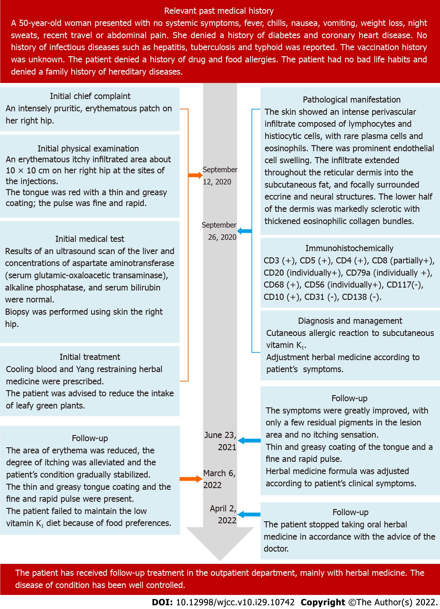
Figure 5 Case timeline.
Important dates, times, and detailed diagnostic process during the treatment and follow-up period.
- Citation: Zhang M, Chen J, Wang CX, Lin NX, Li X. Cutaneous allergic reaction to subcutaneous vitamin K1: A case report and review of literature. World J Clin Cases 2022; 10(29): 10742-10754
- URL: https://www.wjgnet.com/2307-8960/full/v10/i29/10742.htm
- DOI: https://dx.doi.org/10.12998/wjcc.v10.i29.10742


