Copyright
©The Author(s) 2022.
World J Clin Cases. Aug 26, 2022; 10(24): 8797-8804
Published online Aug 26, 2022. doi: 10.12998/wjcc.v10.i24.8797
Published online Aug 26, 2022. doi: 10.12998/wjcc.v10.i24.8797
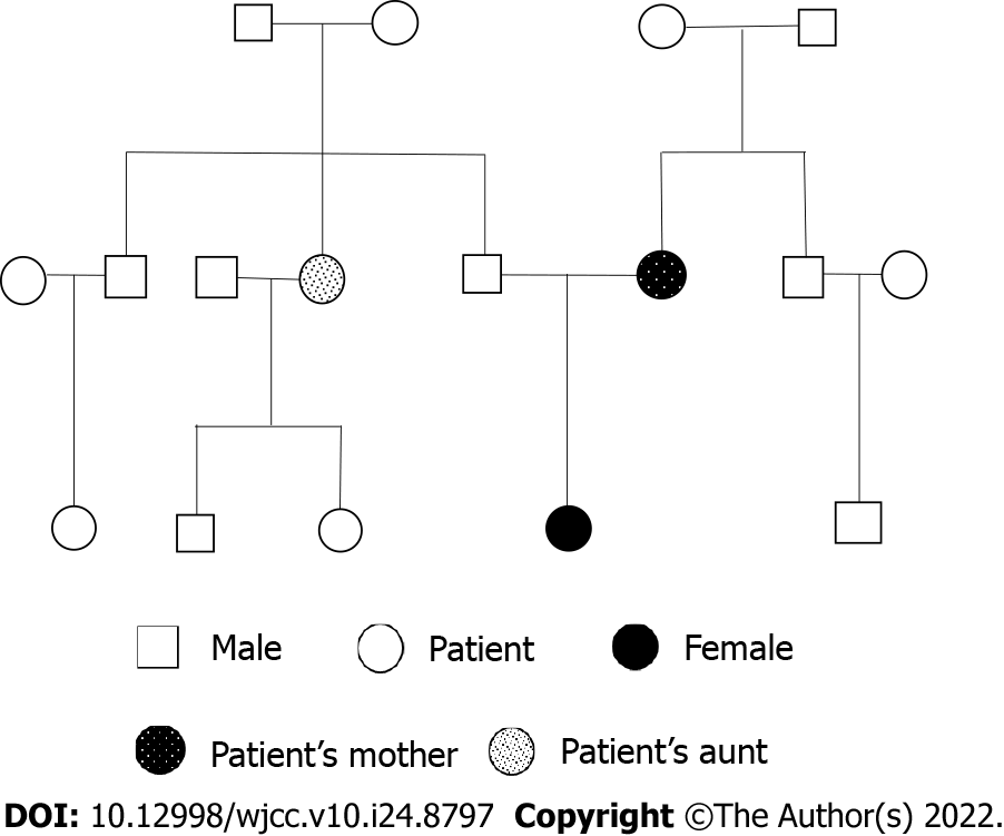
Figure 1 Pedigree diagram of the family.
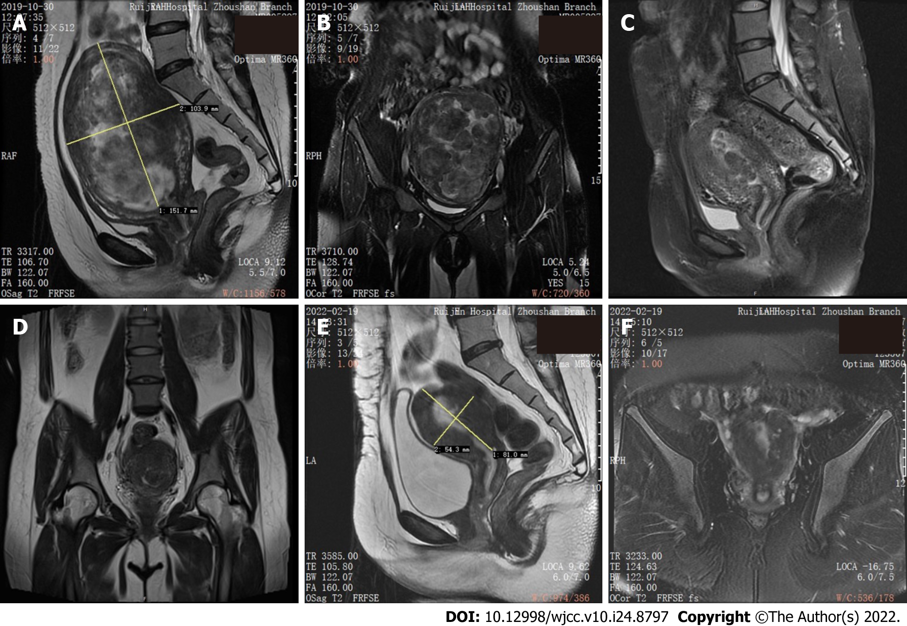
Figure 2 Pelvic magnetic resonance imaging T2-weighted images before and after operations.
A: Sagittal section before transabdominal myomectomy (TM); B: Coronal section before TM; C: Sagittal section before hysteroscopic myomectomy (HM); D: Coronal section before HM; E: Sagittal section after HM; F: Coronal section after HM.
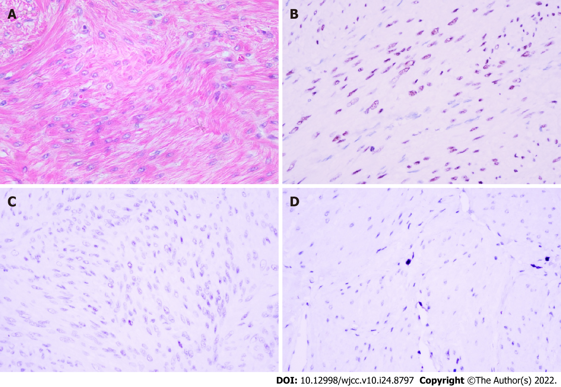
Figure 3 Pathological features of diffuse uterine leiomyomatosis at 400 × original magnification.
A: The lesion displayed typical spindle-shaped smooth muscle cells forming an interlacing fascicular arrangement with ill-defined cell borders, eosinophilic filamentous cytoplasm, cigar-shaped nuclei, and small nucleoli. Mitotic activity was uniformly low (HE staining); B: Immunohistochemistry: Progesterone receptor-positive, 50%-60%; C: Immunohistochemistry: Estrogen receptor-negative; D: Immunohistochemistry: Ki-67-positive, approximately 5%.
- Citation: Ren HM, Wang QZ, Wang JN, Hong GJ, Zhou S, Zhu JY, Li SJ. Diffuse uterine leiomyomatosis: A case report and review of literature. World J Clin Cases 2022; 10(24): 8797-8804
- URL: https://www.wjgnet.com/2307-8960/full/v10/i24/8797.htm
- DOI: https://dx.doi.org/10.12998/wjcc.v10.i24.8797


