Copyright
©The Author(s) 2022.
World J Clin Cases. Aug 26, 2022; 10(24): 8782-8787
Published online Aug 26, 2022. doi: 10.12998/wjcc.v10.i24.8782
Published online Aug 26, 2022. doi: 10.12998/wjcc.v10.i24.8782
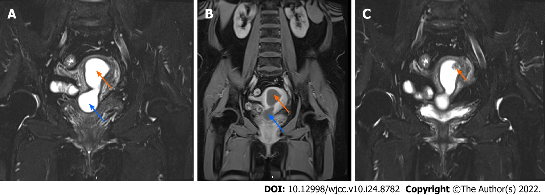
Figure 1 Magnetic resonance imaging of the uterine cavity.
A and B: T2 sequencing (A) and enhanced sequencing (B) of the uterus, both showing fluid within the uterine cavity (orange arrow) and the cervical canal (blue arrow), which was most likely pyometra; C: A mass that protruded into the uterine cavity (arrow), which is most likely an endometrial polyp or submucosal myoma.
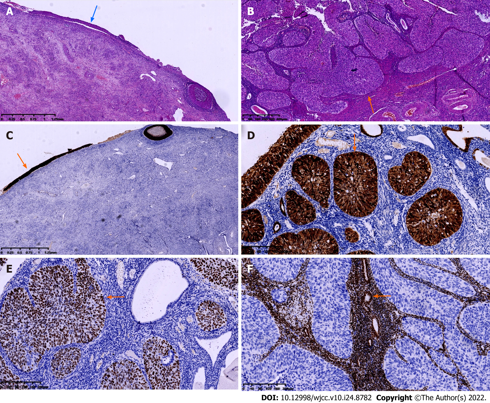
Figure 2 Microscopic appearance of the disease.
A and B: Hematoxylin–eosin staining of the cervix (A) and endometrium (B). While it was a high-grade squamous intraepithelial lesion in the cervix (blue arrow), the lesion penetrated into the endometrium and the myometrium, forming squamous cell carcinoma in the uterine cavity (orange arrow); C-F: Immunohistochemical staining of the uterus. Both the cervix (C) and endometrium (D) showed strong expression of p16 (arrow), p63 was also highly expressed in the endometrium (E), whereas expression of estrogen receptor was negative in the endometrium, and normal endometrial glands (arrow) could also be seen (F).
- Citation: Shu XY, Dai Z, Zhang S, Yang HX, Bi H. Endometrial squamous cell carcinoma originating from the cervix: A case report. World J Clin Cases 2022; 10(24): 8782-8787
- URL: https://www.wjgnet.com/2307-8960/full/v10/i24/8782.htm
- DOI: https://dx.doi.org/10.12998/wjcc.v10.i24.8782


