Copyright
©The Author(s) 2022.
World J Clin Cases. Aug 26, 2022; 10(24): 8728-8734
Published online Aug 26, 2022. doi: 10.12998/wjcc.v10.i24.8728
Published online Aug 26, 2022. doi: 10.12998/wjcc.v10.i24.8728
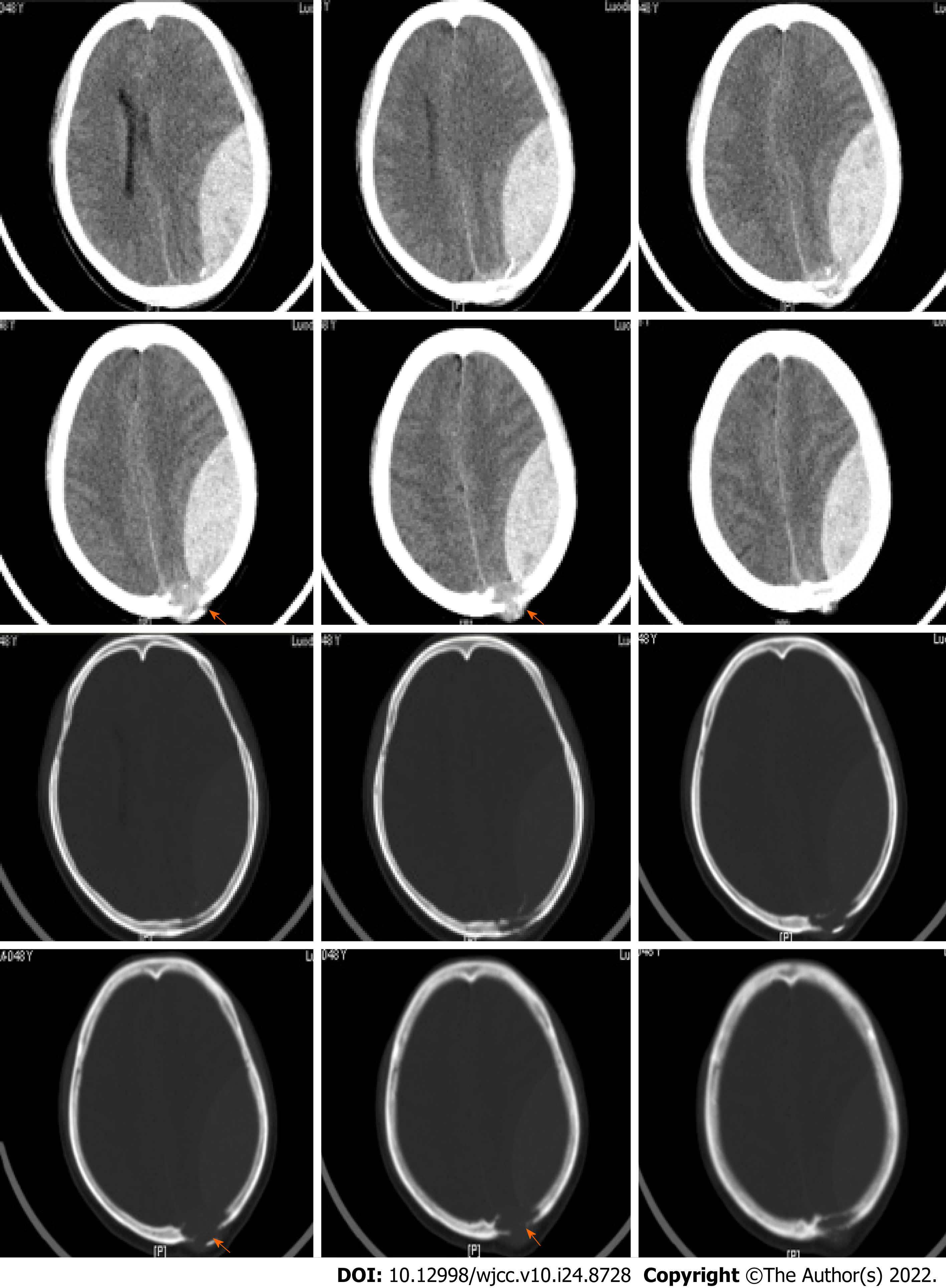
Figure 1 The head computed tomography scan of the patient showed a huge acute epidural hematoma in the left parietal and occipital region.
An osteolytic destruction of the left parietal bone can be found (indicated by the orange arrows).
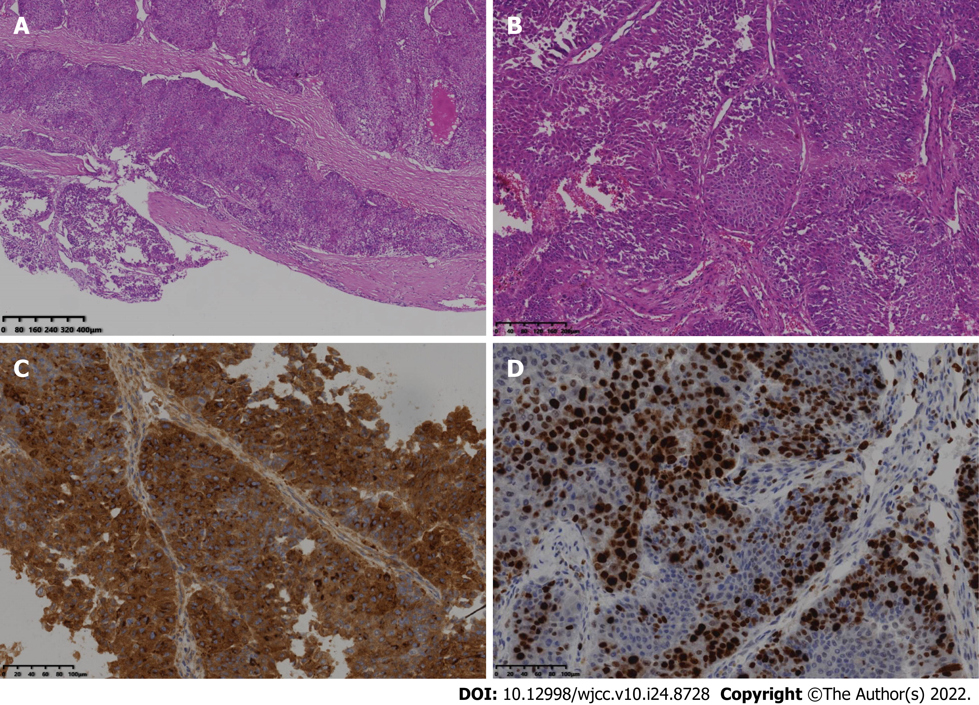
Figure 2 Pathological examination of the lesion.
A: Low-powered picture of the HE staining revealed that the dura mater was invaded by the metastatic tumor; B: High-powered observation of the HE staining showed a sinusoid structure of the metastatic hepatocellular carcinoma. Immuno-histochemistry staining showed that metastatic hepatocellular carcinoma was strongly positive for C: AFP and D: Ki67.
- Citation: Lv GZ, Li GC, Tang WT, Zhou D, Yang Y. Spontaneous acute epidural hematoma secondary to skull and dural metastasis of hepatocellular carcinoma: A case report. World J Clin Cases 2022; 10(24): 8728-8734
- URL: https://www.wjgnet.com/2307-8960/full/v10/i24/8728.htm
- DOI: https://dx.doi.org/10.12998/wjcc.v10.i24.8728


