Copyright
©The Author(s) 2022.
World J Clin Cases. Aug 26, 2022; 10(24): 8656-8661
Published online Aug 26, 2022. doi: 10.12998/wjcc.v10.i24.8656
Published online Aug 26, 2022. doi: 10.12998/wjcc.v10.i24.8656
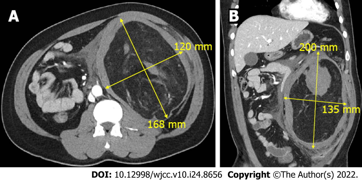
Figure 1 A 30-year-old male patient underwent computed tomography to reveal a 22 cm × 13 cm renal angiomyolipoma.
A: The axial view; B: The coronal view.
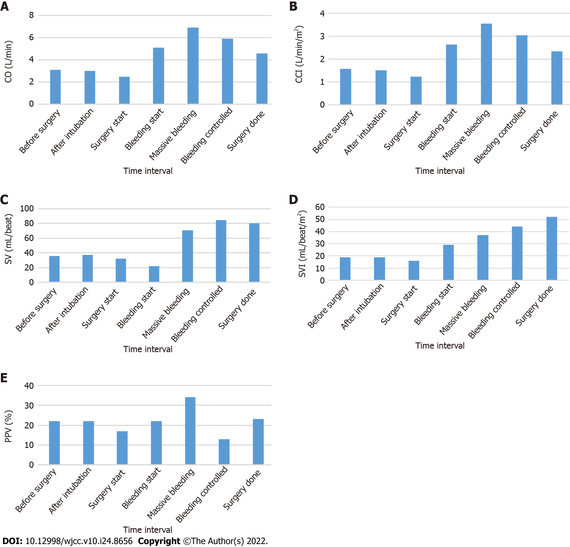
Figure 2 Cardiac parameters were monitored non-invasively using CSN-1901 (Nihon Kohden, Tokyo, Japan) during nephrectomy of a 30-year-old male patients who was diagnosed with ruptured angiomyolipoma.
Additionally, pulse pressure variation was monitored by intra-arterial catheter. A: Cardiac output; B: Continuous cardiac output index; C: Stroke volume; D: Stroke volume index; E: Pulse pressure variation. CO: Cardiac output; CCI: Continuous cardiac index; SV: Stroke volume; SVI: Stroke volume index; PPV: Pulse pressure variation.
- Citation: Jeon WJ, Shin WJ, Yoon YJ, Park CW, Shim JH, Cho SY. Anesthetics management of a renal angiomyolipoma using pulse pressure variation and non-invasive cardiac output monitoring: A case report. World J Clin Cases 2022; 10(24): 8656-8661
- URL: https://www.wjgnet.com/2307-8960/full/v10/i24/8656.htm
- DOI: https://dx.doi.org/10.12998/wjcc.v10.i24.8656


