Copyright
©The Author(s) 2022.
World J Clin Cases. Aug 6, 2022; 10(22): 7931-7935
Published online Aug 6, 2022. doi: 10.12998/wjcc.v10.i22.7931
Published online Aug 6, 2022. doi: 10.12998/wjcc.v10.i22.7931
Figure 1 Computed tomography scan of paraganglioma.
Performed on September 1, 2016: 64-slice computed tomography plain scan + enhanced scan (arrow). A mass of approximately 84 mm × 61 mm (right and left × back and forth) was observed below the left renal artery and vein, the abdominal aorta, the left psoas major muscle and the front of the left kidney. The edge was smooth, with an uneven density. The plain scan computed tomography value was within 17–41 HU. The arrow indicates the location, shape and size of the mass.
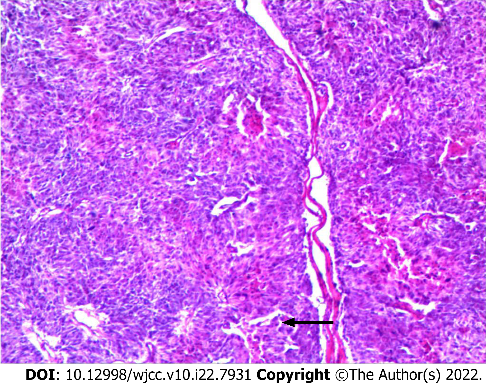
Figure 2 Histopathological features of paraganglioma.
The tumor represents characteristic nest-like structure (arrow, hematoxylin and eosin × 40). The physician who completed the pathological diagnosis was the chief physician, who had been engaged in pathological diagnosis for 31 years. The arrow refers to the typical pathological feature of paraganglioma - nest like structure.
- Citation: Wei JH, Yan HL. Primary hypertension in a postoperative paraganglioma patient: A case report. World J Clin Cases 2022; 10(22): 7931-7935
- URL: https://www.wjgnet.com/2307-8960/full/v10/i22/7931.htm
- DOI: https://dx.doi.org/10.12998/wjcc.v10.i22.7931


