Copyright
©The Author(s) 2022.
World J Clin Cases. Jun 6, 2022; 10(16): 5463-5469
Published online Jun 6, 2022. doi: 10.12998/wjcc.v10.i16.5463
Published online Jun 6, 2022. doi: 10.12998/wjcc.v10.i16.5463
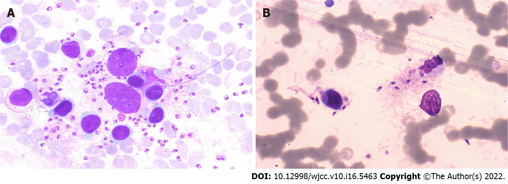
Figure 1 Leishmania amastigotes inside (A) and outside (B) of phagocytes in the bone marrow of Case 1 (Wright-Giemsa, × 1000).
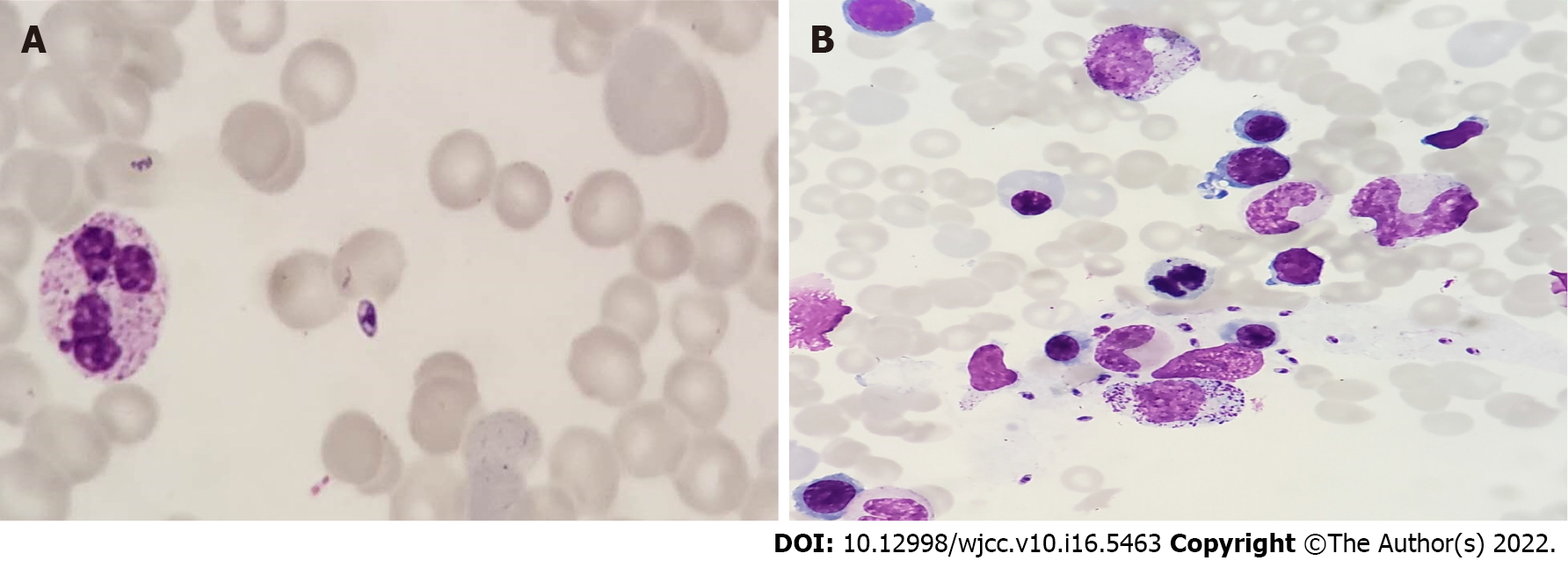
Figure 2 Leishmania amastigotes scattered (A) and piles (B) in the bone marrow of Case 2 (Wright-Giemsa, × 1000).
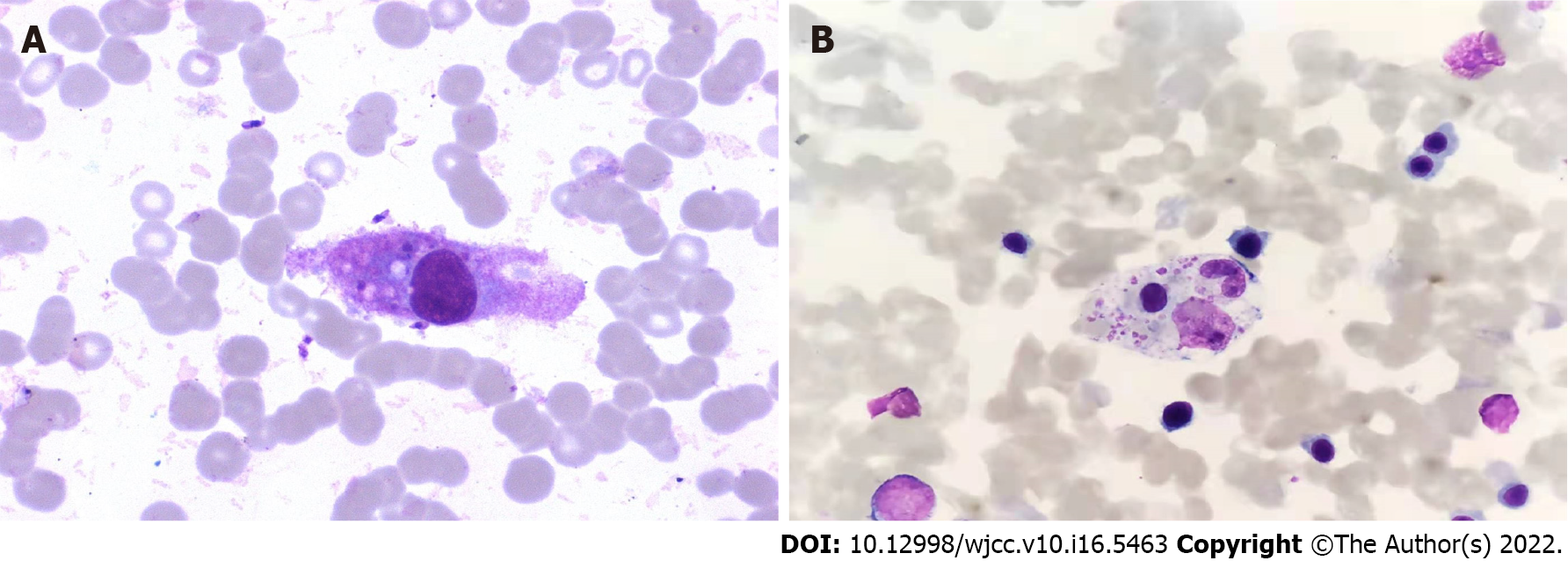
Figure 3 Hemophagocytic cells in the bone marrow (Wright-Giemsa, × 1000).
A: Case 1; B: Case 2.
- Citation: Shi SL, Zhao H, Zhou BJ, Ma MB, Li XJ, Xu J, Jiang HC. Diagnostic value of bone marrow cell morphology in visceral leishmaniasis-associated hemophagocytic syndrome: Two case reports. World J Clin Cases 2022; 10(16): 5463-5469
- URL: https://www.wjgnet.com/2307-8960/full/v10/i16/5463.htm
- DOI: https://dx.doi.org/10.12998/wjcc.v10.i16.5463


