Copyright
©The Author(s) 2022.
World J Clin Cases. Jun 6, 2022; 10(16): 5373-5379
Published online Jun 6, 2022. doi: 10.12998/wjcc.v10.i16.5373
Published online Jun 6, 2022. doi: 10.12998/wjcc.v10.i16.5373
Figure 1 Callosities at the plantar aspect of the heel of the right foot.
A: Lateral position; B: Direct vision; C: Prone position.
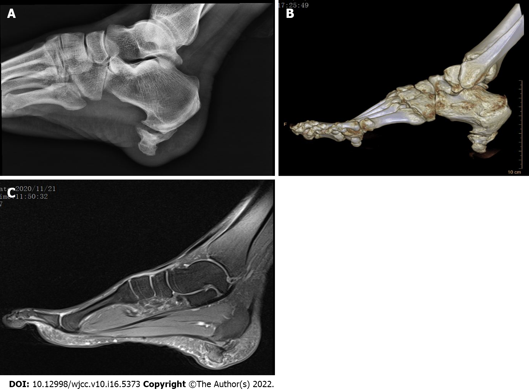
Figure 2 Imaging examinations of the left ankle/calcaneus demonstrated a bony structure on the plantar side of the calcaneus.
A: Radiograph; B: Computer tomography scan; C: Magnetic resonance imaging.
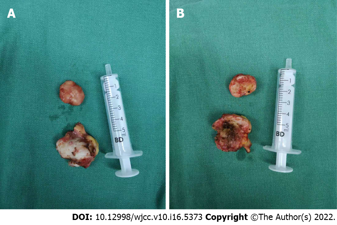
Figure 3 Macroscopic view of the resected os subcalcis.
A: Distal surface; B: Proximal bone surface.
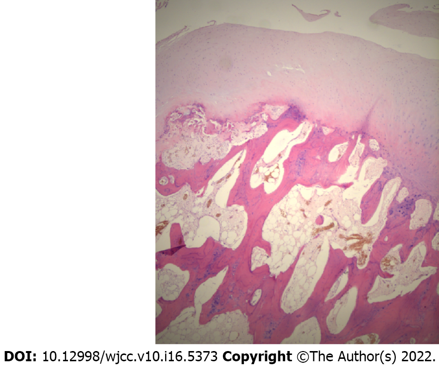
Figure 4
Microscopy showed that the bony trabeculae were intermingled with fat and covered with cartilage.
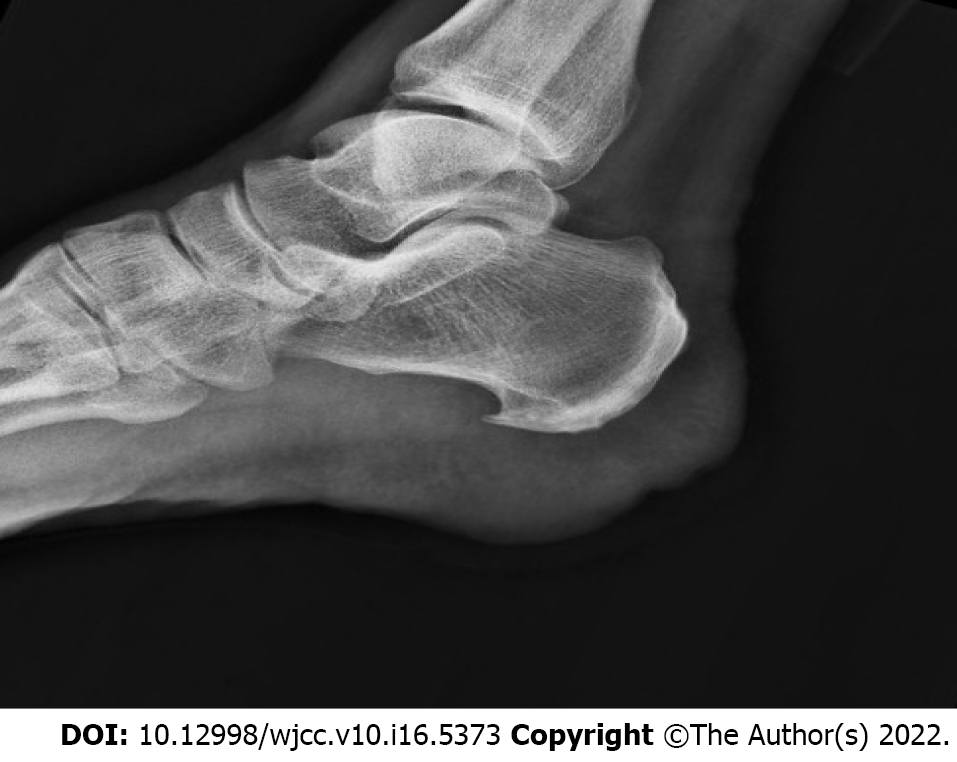
Figure 5
Radiograph at the first postoperative day demonstrated that the os subcalcis was completely resected.
- Citation: Saijilafu, Li SY, Yu X, Li ZQ, Yang G, Lv JH, Chen GX, Xu RJ. Heel pain caused by os subcalcis: A case report. World J Clin Cases 2022; 10(16): 5373-5379
- URL: https://www.wjgnet.com/2307-8960/full/v10/i16/5373.htm
- DOI: https://dx.doi.org/10.12998/wjcc.v10.i16.5373


