Copyright
©The Author(s) 2022.
World J Clin Cases. Jun 6, 2022; 10(16): 5324-5330
Published online Jun 6, 2022. doi: 10.12998/wjcc.v10.i16.5324
Published online Jun 6, 2022. doi: 10.12998/wjcc.v10.i16.5324
Figure 1 Computed tomography on admission.
A and C: Horizontal section; B: Coronal section. Computed tomography revealed a soft tissue mass dorsal to the stomach (C: white dotted line) and nearby duodenojejunal flexure (A and B: white arrow head). Dilated duodenum (d) and collapsed jejunum (A and B: white arrow) was found. g: Stomach; d: Duodenum; p: Pancreas.
Figure 2 Preoperative upper gastrointestinal investigation and upper gastrointestinal endoscopy.
A: Upper gastrointestinal investigation showed a dilated duodenum (d), stomach lacking extensibility (g) and no gastrografin passage through the duodenojejunal flexure (white arrow); B: Upper gastrointestinal endoscopy could not pass through the duodenojejunal flexure due to intraluminal stenosis (white arrow) but revealed no mucosal surface change; C: A nasogastric tube was placed to decompress the stomach and duodenum. g: Stomach; d: Duodenum.
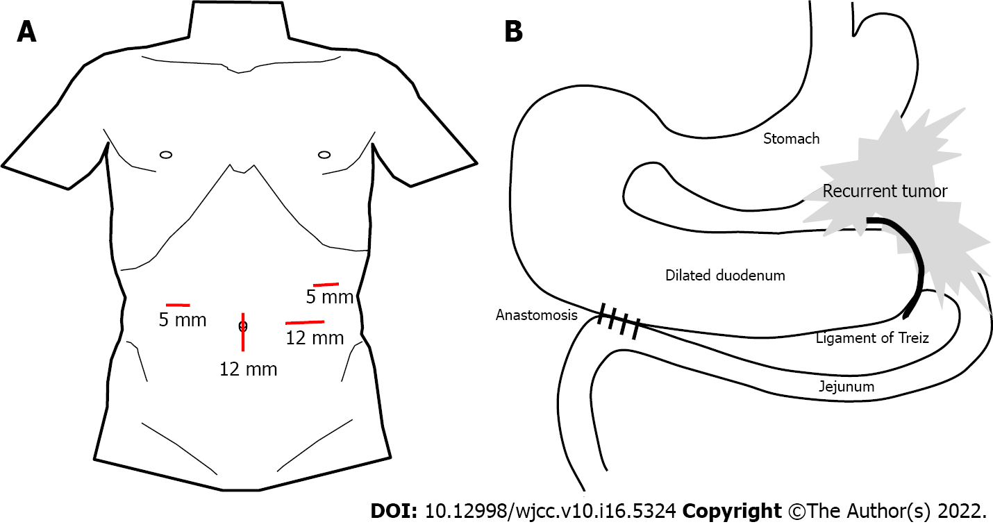
Figure 3 Schematic illustration.
A: Port placement; B: Anatomy of anastomosis in laparoscopic duodenojejunostomy.
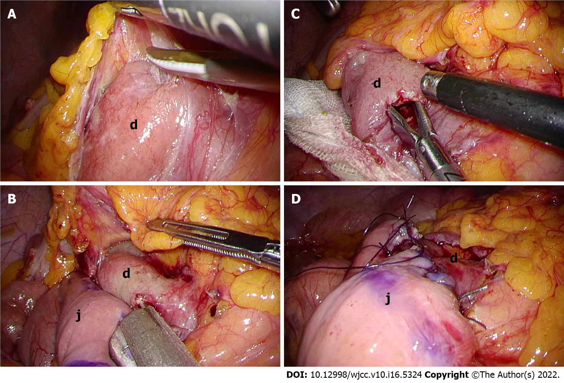
Figure 4 Images of duodenojejunostomy.
A: The second and third portions of the duodenum (d) were exposed and mobilized; B: Enterotomy was created in the third potion of the duodenum (d) and jejunum (j) about 30 cm anal to the Treitz ligament for anastomosis; C: A side-to-side duodenojejunostomy was performed in the manner of antiperistalsis using 45-mm stapling device; D: The common entry hole was closed with a continuous suture. g: Stomach; j: Jejunum.
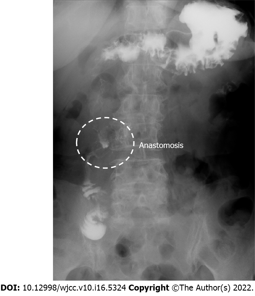
Figure 5 Upper gastrointestinal investigation after laparoscopic duodenojejunostomy.
Gastrografin passed from the duodenum into the jejunum through the anastomosis.
- Citation: Murakami T, Matsui Y. Laparoscopic duodenojejunostomy for malignant stenosis as a part of multimodal therapy: A case report. World J Clin Cases 2022; 10(16): 5324-5330
- URL: https://www.wjgnet.com/2307-8960/full/v10/i16/5324.htm
- DOI: https://dx.doi.org/10.12998/wjcc.v10.i16.5324


