Copyright
©The Author(s) 2022.
World J Clin Cases. Jun 6, 2022; 10(16): 5317-5323
Published online Jun 6, 2022. doi: 10.12998/wjcc.v10.i16.5317
Published online Jun 6, 2022. doi: 10.12998/wjcc.v10.i16.5317
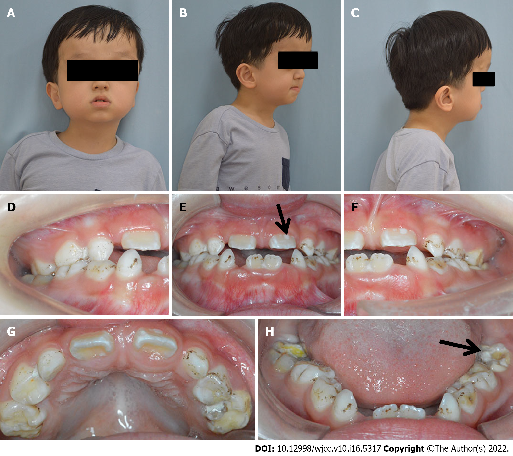
Figure 1 Extraoral and intraoral presentations of the patient.
A-C: Extraoral photos showed a prominent forehead, ocular proptosis, midface hypoplasia, retrusive upper lip, and protrusive lower lip; D-H: The intraoral photos showed that oral hygiene was poor with scattered pigmentation on the teeth. Teeth 36, 46, 54, 55, 65, 74, 75, and 85 were decayed, and teeth 11, 21, 36, and 46 showed hypomineralization (black arrows).
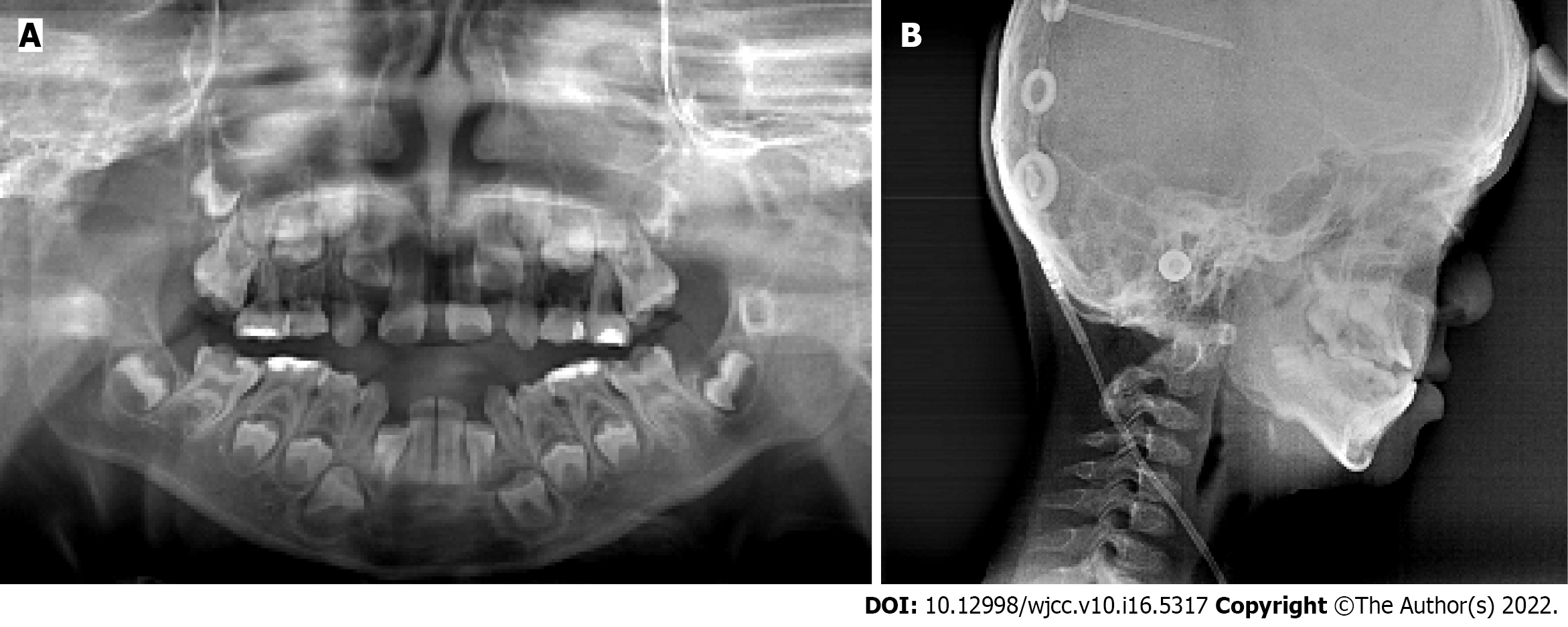
Figure 2 Panoramic and lateral cephalometric radiograph of the patient.
A: The panoramic radiograph showed normal permanent tooth germ development; B: The lateral cephalometric radiograph showed a skeletal Class III relationship.
- Citation: Li XJ, Su JM, Ye XW. Crouzon syndrome in a fraternal twin: A case report and review of the literature. World J Clin Cases 2022; 10(16): 5317-5323
- URL: https://www.wjgnet.com/2307-8960/full/v10/i16/5317.htm
- DOI: https://dx.doi.org/10.12998/wjcc.v10.i16.5317


