Copyright
©The Author(s) 2022.
World J Clin Cases. May 26, 2022; 10(15): 5025-5035
Published online May 26, 2022. doi: 10.12998/wjcc.v10.i15.5025
Published online May 26, 2022. doi: 10.12998/wjcc.v10.i15.5025
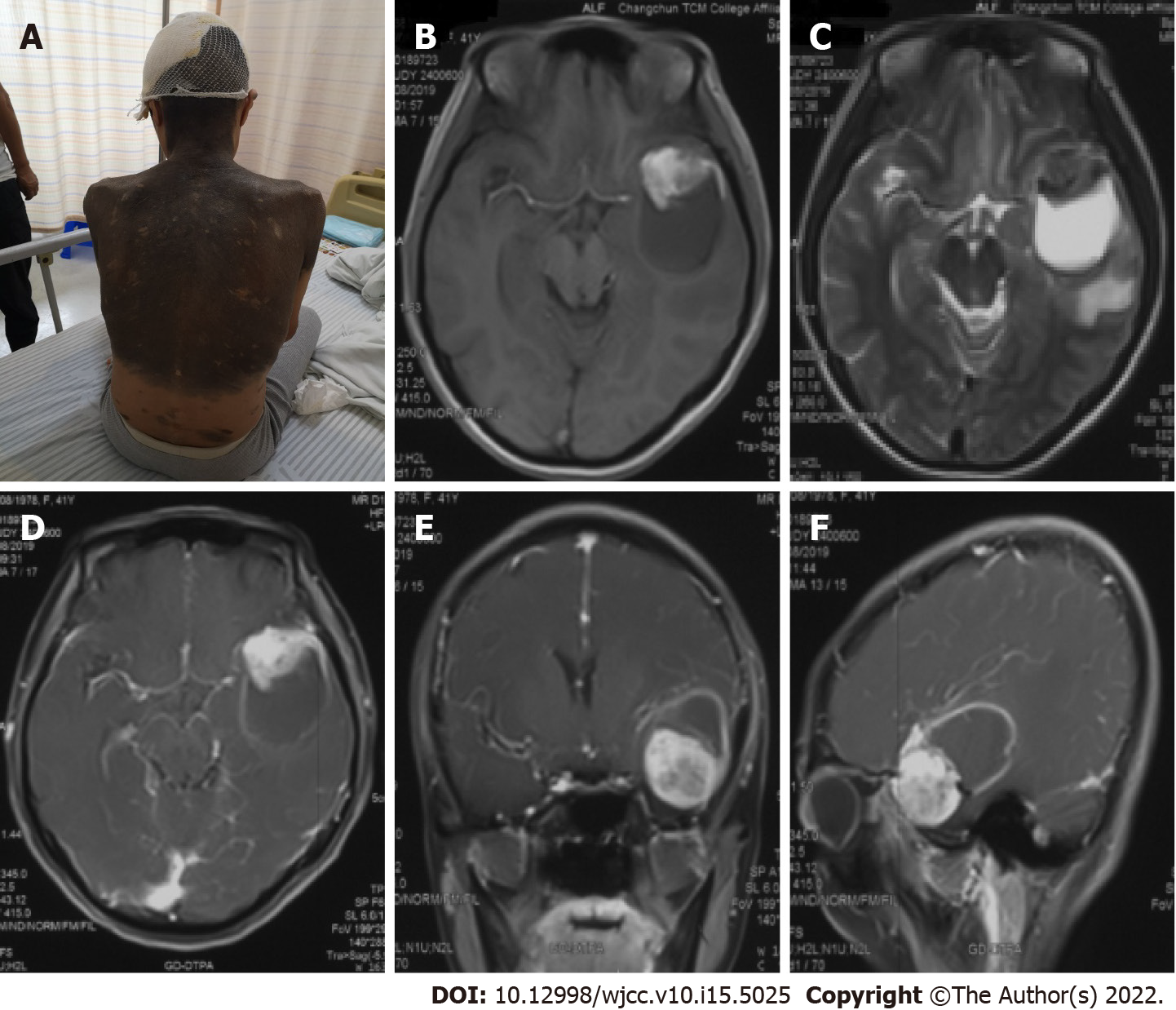
Figure 1 Images and physical examination.
A: Physical examination revealed a giant skin nevus; B: T1-weighted; C: T2-weighted; D-F: After the injection of a contrast agent.
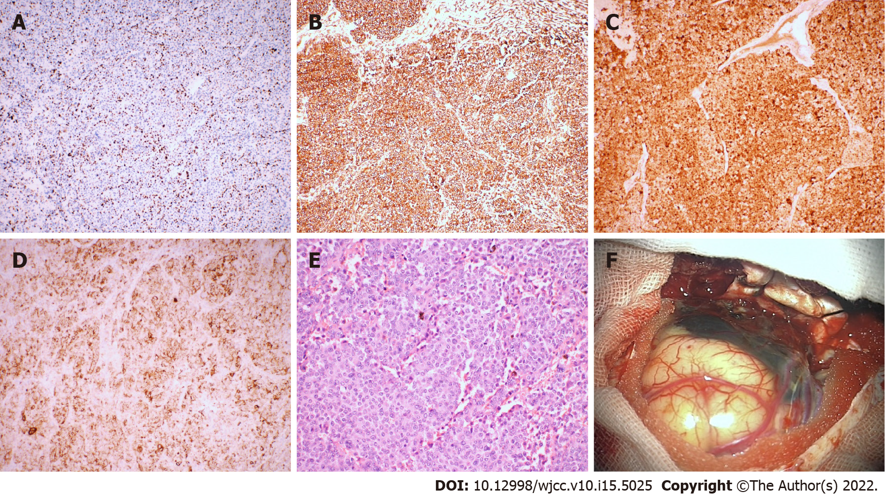
Figure 2 Immunohistochemistry and direct observation of the tumor.
A: Ki-67 (+30%); B: Vimentin (+); C: S-100 (+); D: HMB45 (+); E: × 200, HE; F: The tumor appeared blackish brown, had no boundary or capsule, and was soft; the peripheral leptomeninges were also stained black.
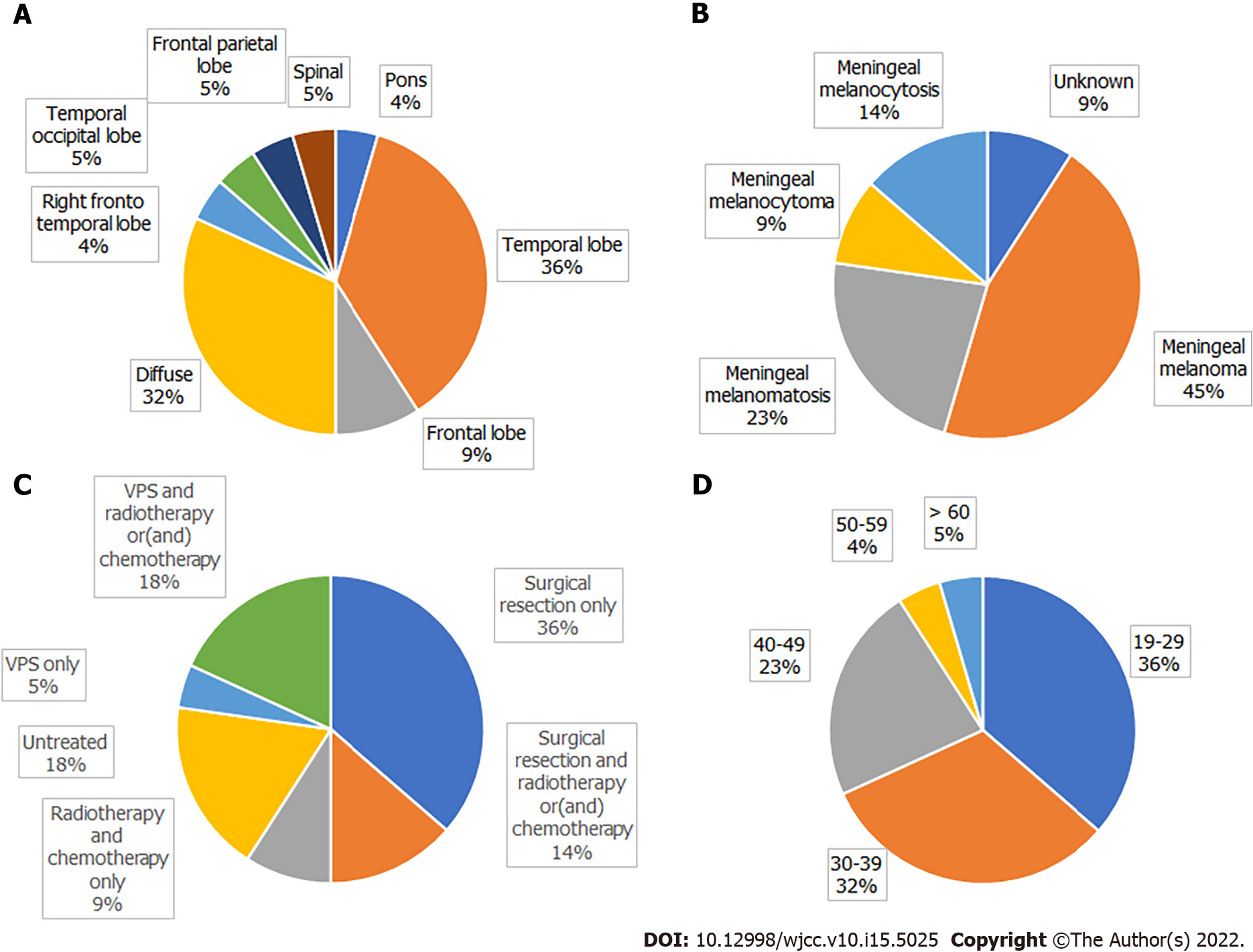
Figure 3 Distribution of patients.
A: The location of the tumor; B: Pathological type; C: Treatment; D: Age.
- Citation: Liu BC, Wang YB, Liu Z, Jiao Y, Zhang XF. Neurocutaneous melanosis with an intracranial cystic-solid meningeal melanoma in an adult: A case report and review of literature. World J Clin Cases 2022; 10(15): 5025-5035
- URL: https://www.wjgnet.com/2307-8960/full/v10/i15/5025.htm
- DOI: https://dx.doi.org/10.12998/wjcc.v10.i15.5025


