Copyright
©The Author(s) 2022.
World J Clin Cases. May 26, 2022; 10(15): 4957-4963
Published online May 26, 2022. doi: 10.12998/wjcc.v10.i15.4957
Published online May 26, 2022. doi: 10.12998/wjcc.v10.i15.4957
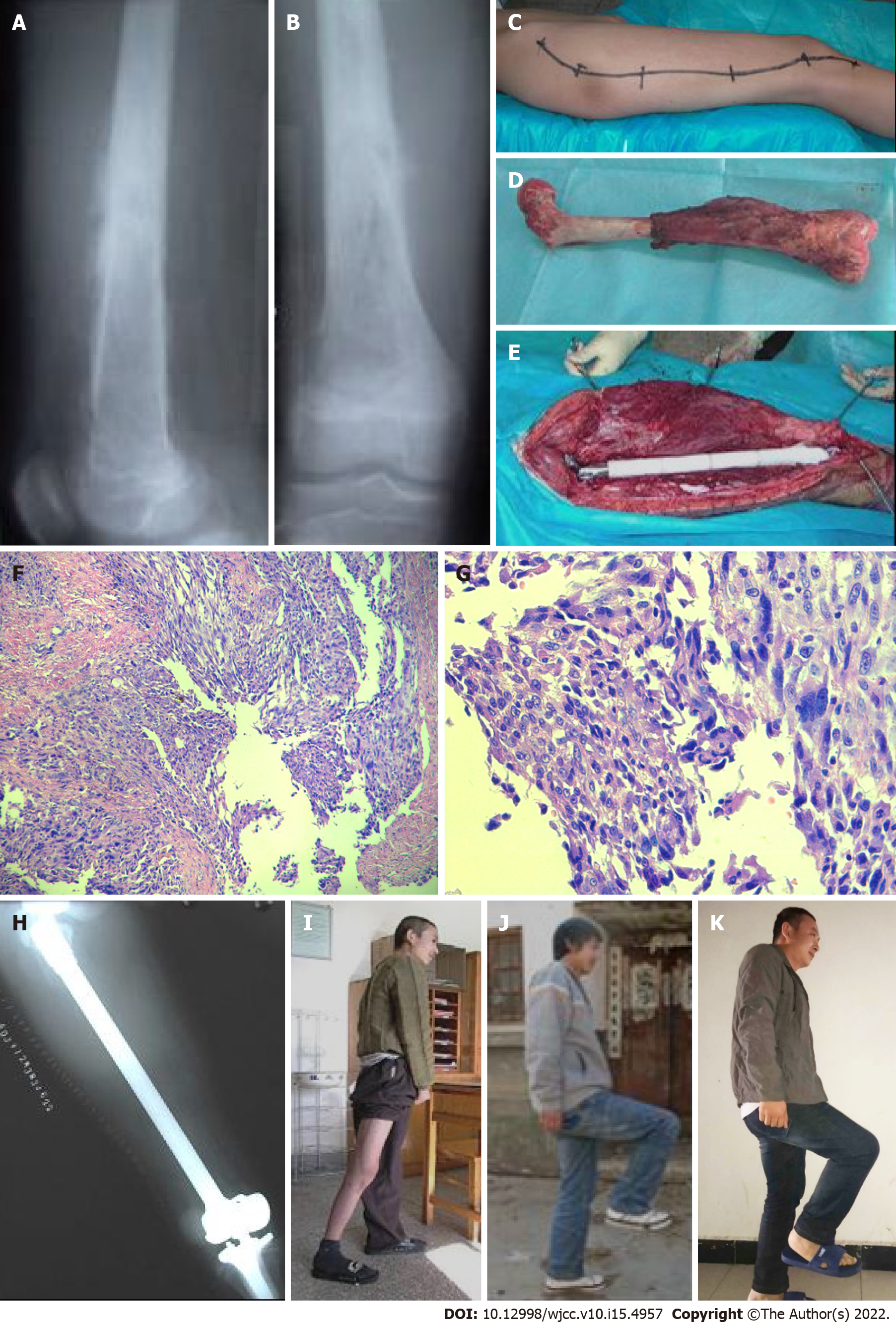
Figure 1 Imaging examinations.
A and B: X-ray illustrates osteolytic destruction in the middle and lower segments of the right femur, with significant periosteal reaction; C-E: Intraoperative condition; F-G: 4×10, 40×10 magnification photomicrograph, respectively, hematoxylin and eosin; H: Postoperative X-ray; I-K: Postoperative follow up after the 1, 4 and 18 years.
- Citation: Yang YH, Chen JX, Chen QY, Wang Y, Zhou YB, Wang HW, Yuan T, Sun HP, Xie L, Yao ZH, Yang ZZ. Total femur replacement with 18 years of follow-up: A case report. World J Clin Cases 2022; 10(15): 4957-4963
- URL: https://www.wjgnet.com/2307-8960/full/v10/i15/4957.htm
- DOI: https://dx.doi.org/10.12998/wjcc.v10.i15.4957


