Copyright
©The Author(s) 2022.
World J Clin Cases. May 16, 2022; 10(14): 4617-4624
Published online May 16, 2022. doi: 10.12998/wjcc.v10.i14.4617
Published online May 16, 2022. doi: 10.12998/wjcc.v10.i14.4617
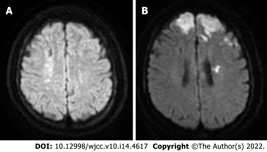
Figure 1 Magnetic resonance imaging of the head during acute cerebral infarction.
A: Magnetic resonance imaging of the patient 4 mo earlier that showed an acute cerebral infarction; B: Acute recurrent cerebral infarction.
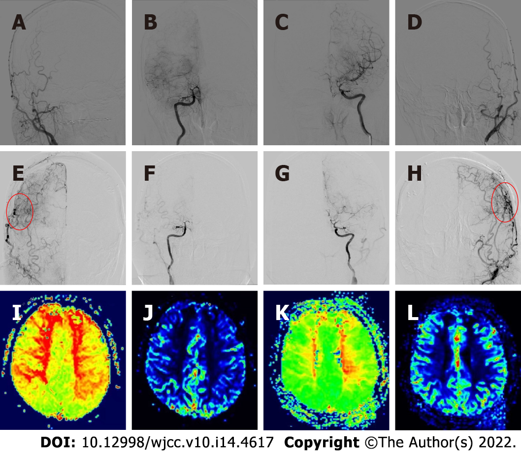
Figure 2 Preoperative and postoperative angiography and cerebral perfusion contrast.
A-D: Preoperative digital subtraction angiography showed moyamoya disease; E-H: The neovascularization was good after bilateral combined cerebral revascularization; I and J: Preoperative magnetic resonance-perfusion-weighted imaging (MR-PWI) showed prolonged time to peak and decreased regional cerebral blood flow in the bilateral frontal parietal lobe, bilateral paraventricular region, and center of semicovale; K and L: Six months after bilateral cerebral revascularization, MR-PWI showed significant improvement in bilateral cerebral perfusion.
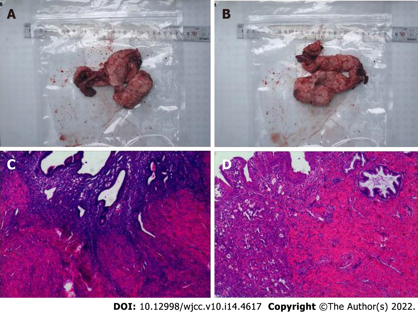
Figure 3 The histopathological examination of the whole uterus and bilateral salpingectomy confirmed adenomyosis.
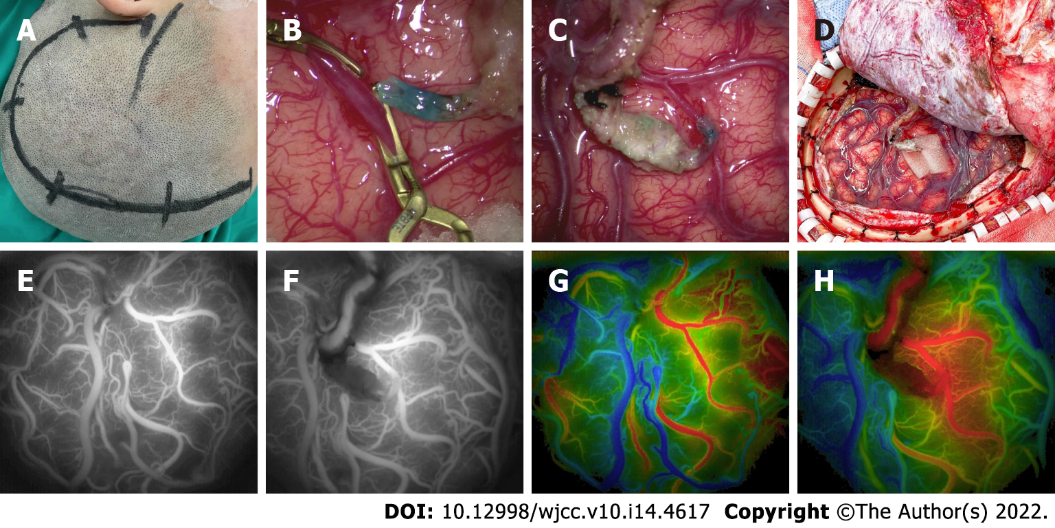
Figure 4 Operative procedure and intraoperative angiography.
A-D: Left-side combined cerebral revascularization surgery; E-H: According to the intraoperative indocyanine green video angiography and Flow 800 technology, the optimal recipient vessels were selected, and the analysis results showed good patency of bridge vessels and improved local cerebral perfusion after bypass surgery.
- Citation: Zhang S, Zhao LM, Xue BQ, Liang H, Guo GC, Liu Y, Wu RY, Li CY. Acute recurrent cerebral infarction caused by moyamoya disease complicated with adenomyosis: A case report . World J Clin Cases 2022; 10(14): 4617-4624
- URL: https://www.wjgnet.com/2307-8960/full/v10/i14/4617.htm
- DOI: https://dx.doi.org/10.12998/wjcc.v10.i14.4617


