Copyright
©The Author(s) 2022.
World J Clin Cases. May 16, 2022; 10(14): 4608-4616
Published online May 16, 2022. doi: 10.12998/wjcc.v10.i14.4608
Published online May 16, 2022. doi: 10.12998/wjcc.v10.i14.4608
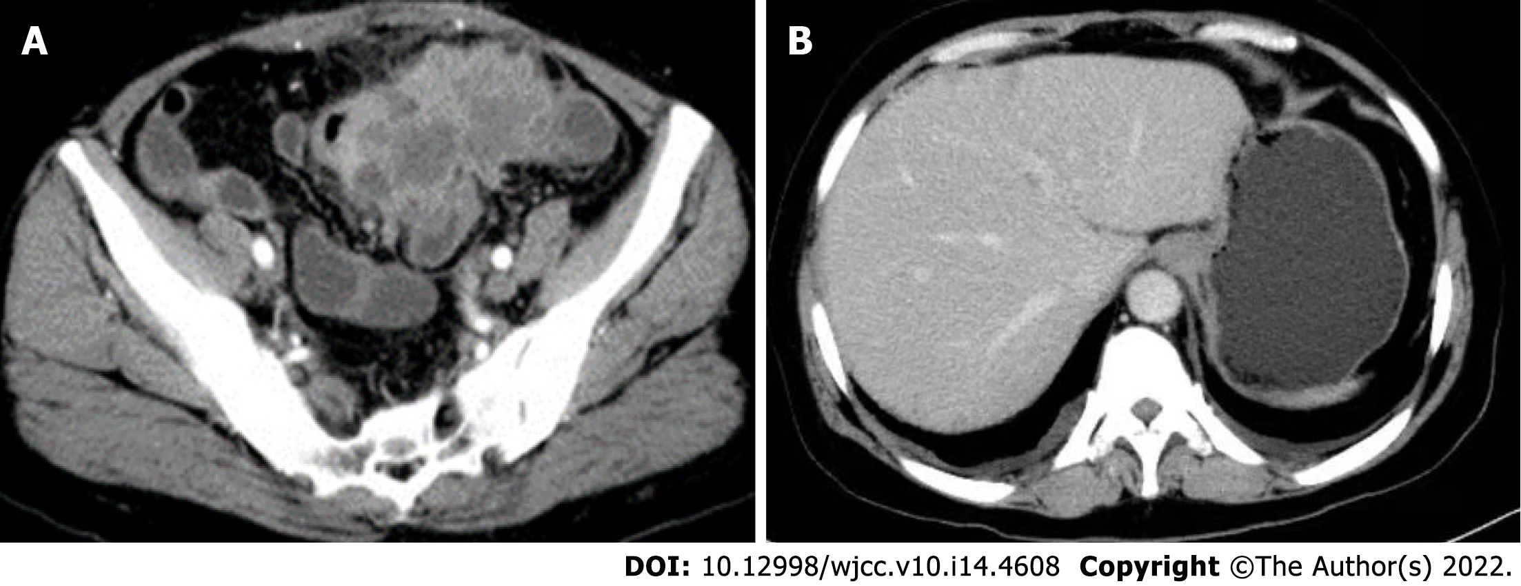
Figure 1 Preoperative computed tomography enhancement scan of the abdomen.
A: An irregular soft tissue mass located in the left lower abdomen with unclear borders and nonuniform density; B: No metastatic lesions in the liver.
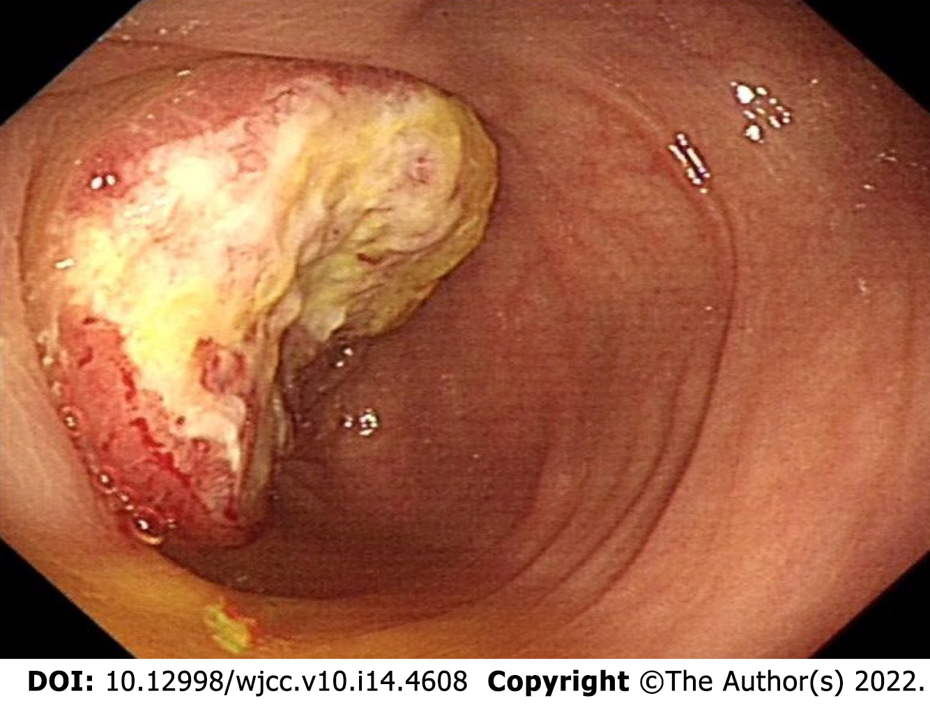
Figure 2 Colonoscopy showing an ulcerated mass with infiltrative growth observed in the colon approximately 30 cm from the anus.
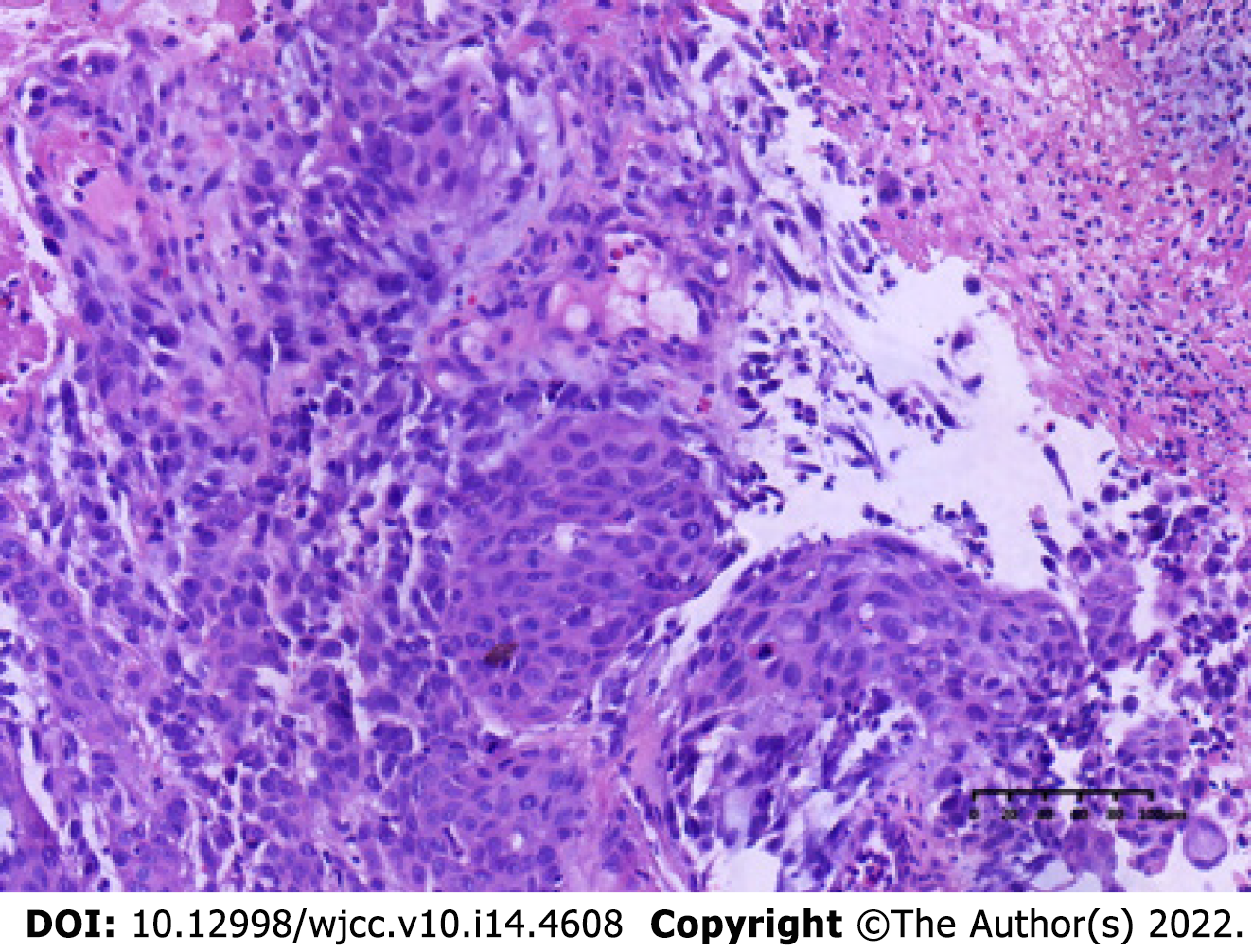
Figure 3 Hematoxylin and eosin staining of tissue specimens taken for colonoscopy.
Anisocytic tumor cells in a striated or scattered pattern (hematoxylin and eosin, 20 ×).
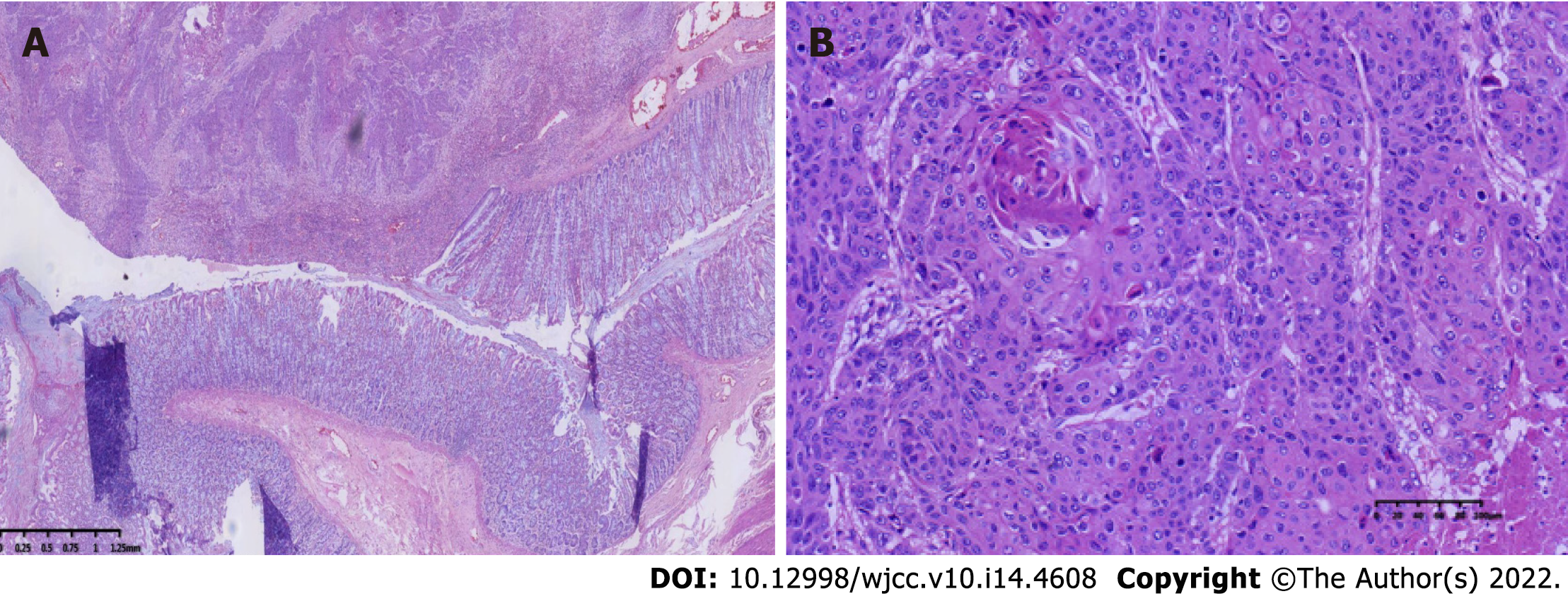
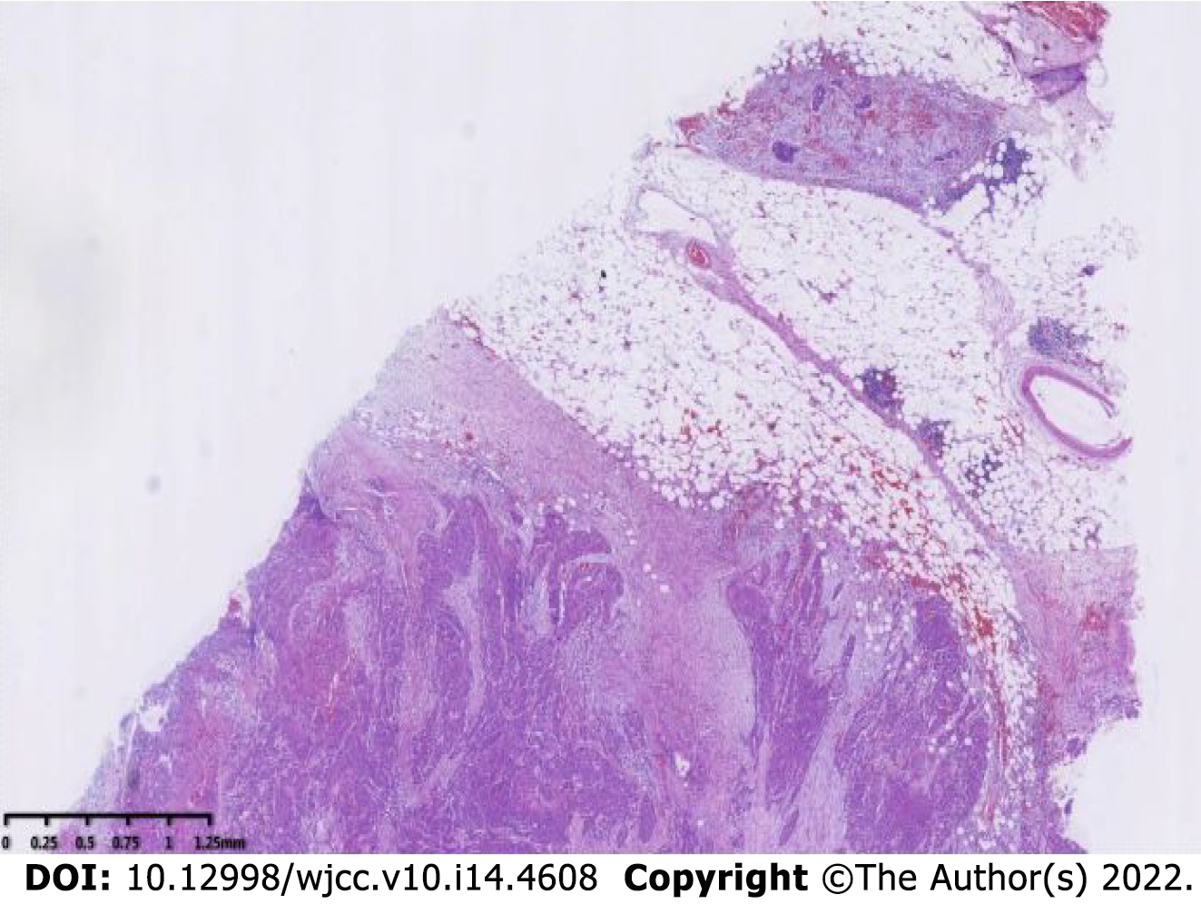
Figure 4 Hematoxylin and eosin staining of tissue specimens taken for surgery.
A: Normal structure and canceration of the sigmoid colon (hematoxylin and eosin, 2 ×); B: Postoperative pathological image (hematoxylin and eosin, 20 ×).
Figure 5 The cancer tissue invades the full thickness of the intestinal wall (hematoxylin and eosin, 2 ×).
Figure 6 Immunohistochemical staining of CDX2, CK5/CK6, and P40.
A: The cancer cells were negative for CDX2 [immunohistochemistry (IHC), 20 ×]; B: The cancer cells were positive for CK5/CK6 (IHC, 20 ×); C: The cancer cells were positive for P40 (IHC, 20 ×).
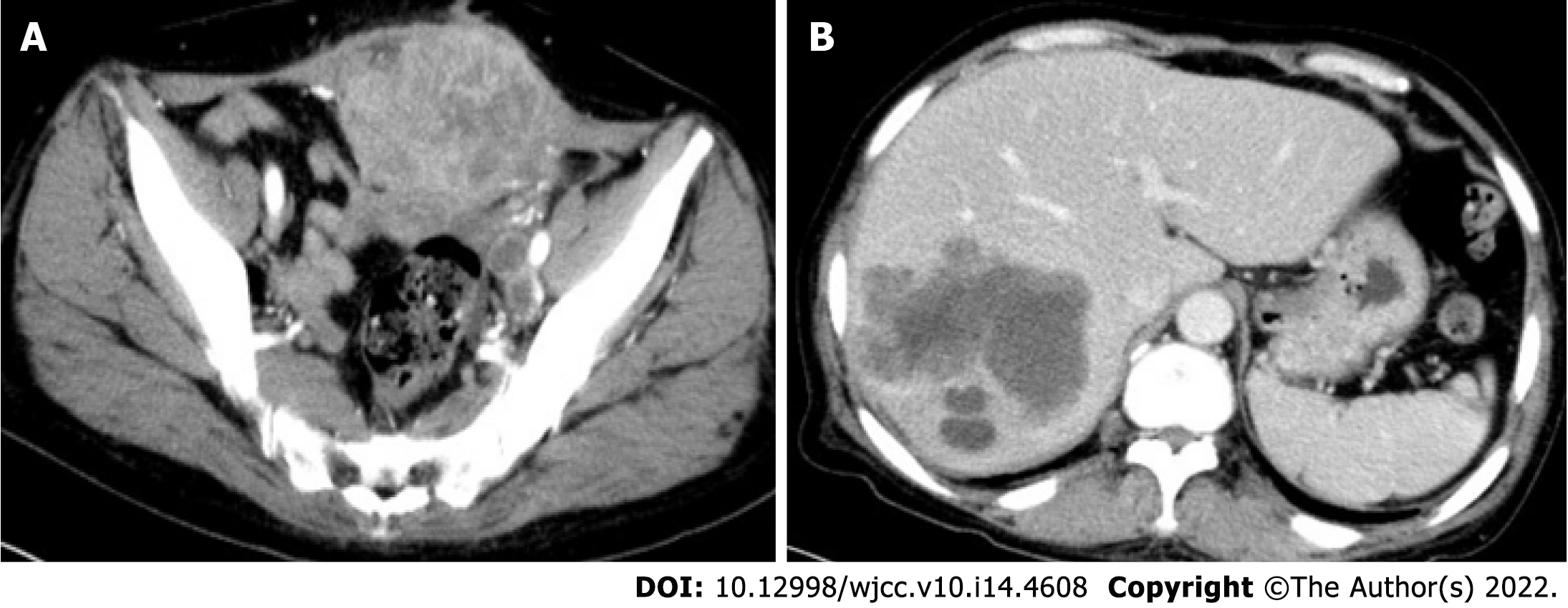
Figure 7 Computed tomography enhancement scan of the abdomen 7 mo postoperatively.
A: Irregular soft tissue mass and nodular shadow were observed in the anterior pelvis, with inhomogeneous enhancement, posterior downward extrusion of the bladder, and indistinct demarcation with the anterior peritoneum and muscles; B: Multiple nodules of variable sizes and mass-like hypointense shadow were observed in the liver.
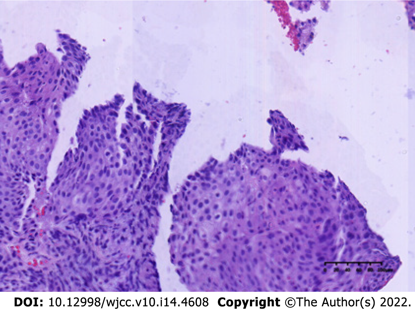
Figure 8 Metastatic squamous cell carcinoma from the liver (hematoxylin and eosin, 20 ×).
- Citation: Li XY, Teng G, Zhao X, Zhu CM. Primary sigmoid squamous cell carcinoma with liver metastasis: A case report. World J Clin Cases 2022; 10(14): 4608-4616
- URL: https://www.wjgnet.com/2307-8960/full/v10/i14/4608.htm
- DOI: https://dx.doi.org/10.12998/wjcc.v10.i14.4608


