Copyright
©The Author(s) 2022.
World J Clin Cases. May 16, 2022; 10(14): 4414-4424
Published online May 16, 2022. doi: 10.12998/wjcc.v10.i14.4414
Published online May 16, 2022. doi: 10.12998/wjcc.v10.i14.4414
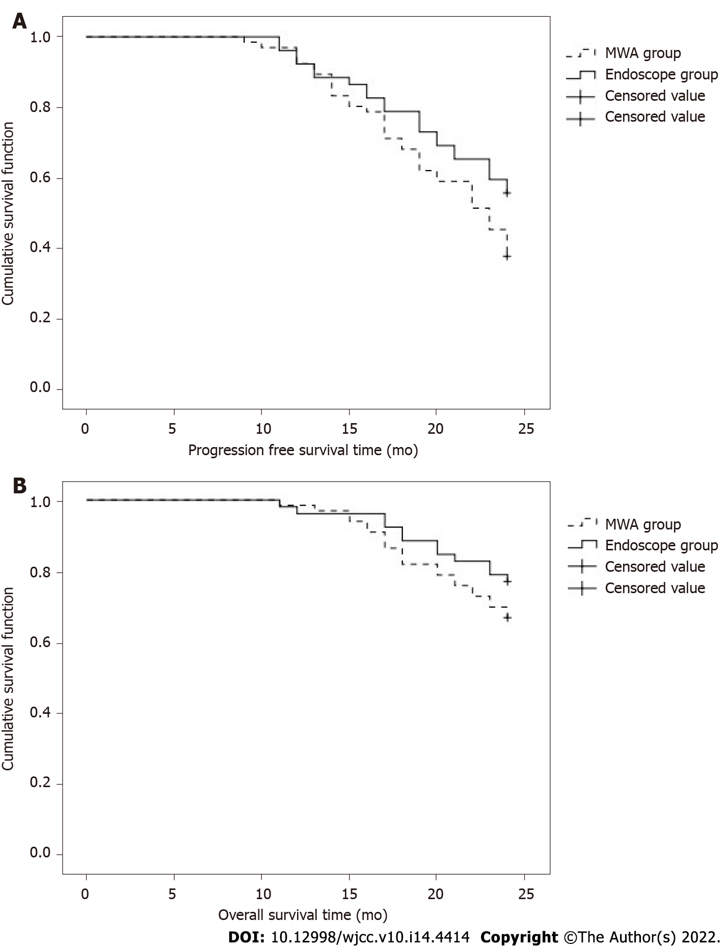
Figure 1 Survival function of the two groups of patients.
A: Progression-free survival function; B: Survival function. MWA: Microwave ablation.
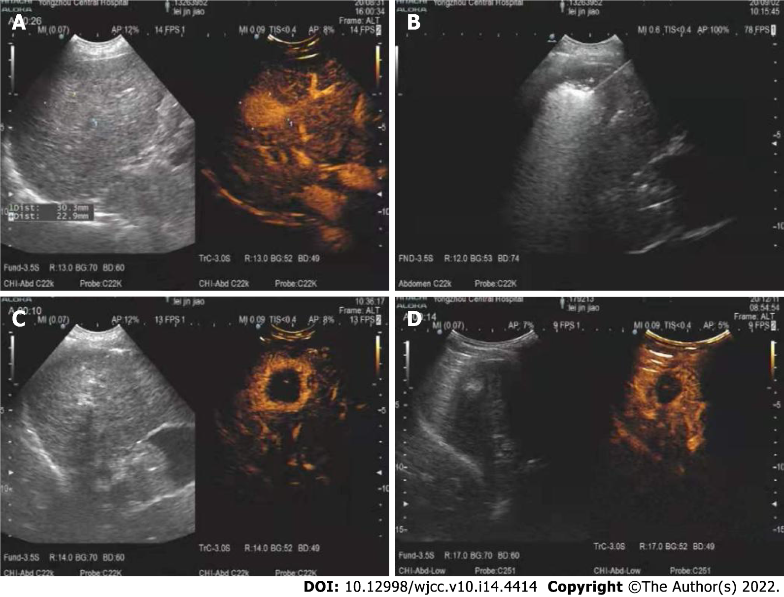
Figure 2 A 75-year-old woman was diagnosed with a right hepatic lobe tumor on preoperative examination.
A: The size of the right hepatic lobe tumor before microwave ablation, with relatively clear boundaries; B: The puncture and ablation process during microwave ablation, indicating that the needle insertion position was relatively accurate; C: The examination after microwave ablation. It can be seen that there is no enhancement in the venous and arterial phases, and the ablation range is 41 cm × 36 cm; D: The reexamination with contrast-enhanced ultrasound 3 mo after ablation, with no enhancement in arterial and venous phases.
- Citation: Zhong H, Hu R, Jiang YS. Evaluation of short- and medium-term efficacy and complications of ultrasound-guided ablation for small liver cancer. World J Clin Cases 2022; 10(14): 4414-4424
- URL: https://www.wjgnet.com/2307-8960/full/v10/i14/4414.htm
- DOI: https://dx.doi.org/10.12998/wjcc.v10.i14.4414


