Copyright
©The Author(s) 2021.
World J Gastrointest Oncol. Jun 15, 2021; 13(6): 472-494
Published online Jun 15, 2021. doi: 10.4251/wjgo.v13.i6.472
Published online Jun 15, 2021. doi: 10.4251/wjgo.v13.i6.472
Figure 1 Hematoxylin and eosin stains[21].
A: Hematoxylin and eosin stains of normal and adjacent ductal adenocarcinoma 40 ×; B: demonstrates invasive adenocarcinoma (100 ×); C: Perineural invasion is demonstrated. Citation: Porta M, Fabregat X, Malats N, Guarner L, Carrato A, de Miguel A, Ruiz L, Jariod M, Costafreda S, Coll S, Alguacil J, Corominas JM, Solà R, Salas A, Real FX. Exocrine pancreatic cancer: symptoms at presentation and their relation to tumour site and stage. Clin Transl Oncol 2005; 7: 189-197. Copyright© The Authors 2005. Published by Springer Nature.
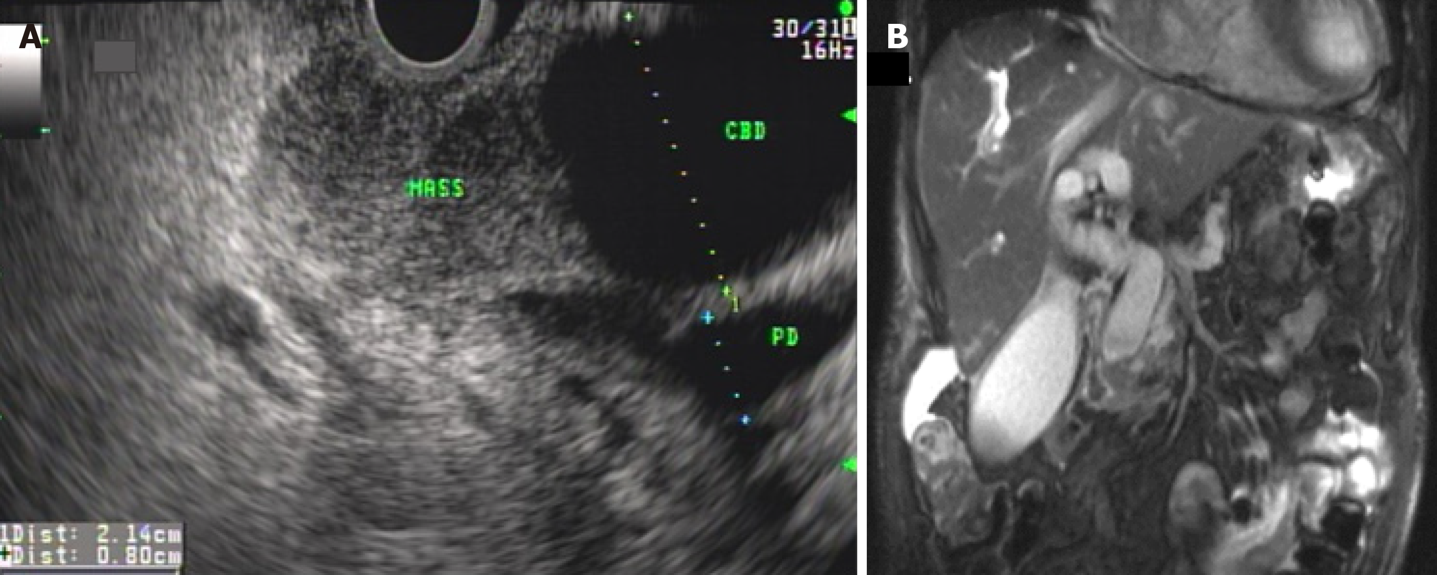
Figure 2 Malignant biliary obstruction from mass in the head of pancreas causing common bile duct and pancreatic duct dilation (double-duct sign) on endoscopic ultrasound and magnetic resonance cholangiopancreatography[21].
A: Endoscopic ultrasound; B: Magnetic resonance cholangiopancreatography, note the distended gallbladder, seen in patients with malignant biliary obstruction (Courvoisier’s sign). Citation: Porta M, Fabregat X, Malats N, Guarner L, Carrato A, de Miguel A, Ruiz L, Jariod M, Costafreda S, Coll S, Alguacil J, Corominas JM, Solà R, Salas A, Real FX. Exocrine pancreatic cancer: symptoms at presentation and their relation to tumour site and stage. Clin Transl Oncol 2005; 7: 189-197. Copyright© The Authors 2005. Published by Springer Nature.
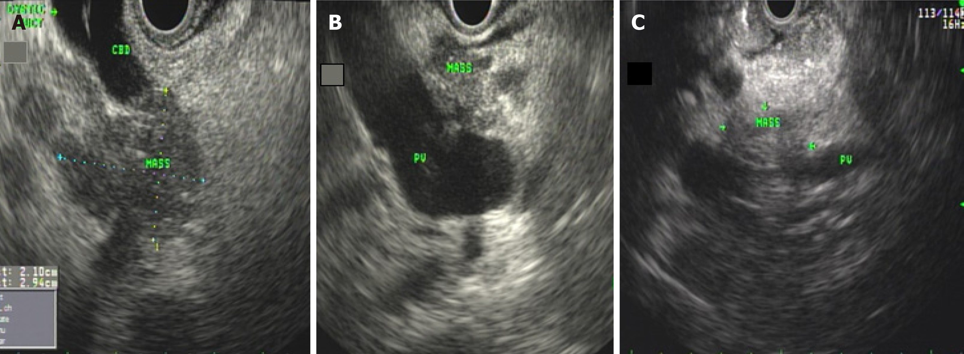
Figure 3 Endoscopic ultrasound images[21].
A: Endoscopic ultrasound (EUS) images of pancreatic adenocarcinoma invading the distal common bile duct; B: EUS images of pancreatic adenocarcinoma invading the portal vein; C: EUS images of pancreatic adenocarcinoma invading the portal vein confluence. Citation: Porta M, Fabregat X, Malats N, Guarner L, Carrato A, de Miguel A, Ruiz L, Jariod M, Costafreda S, Coll S, Alguacil J, Corominas JM, Solà R, Salas A, Real FX. Exocrine pancreatic cancer: symptoms at presentation and their relation to tumour site and stage. Clin Transl Oncol 2005; 7: 189-197. Copyright© The Authors 2005. Published by Springer Nature.
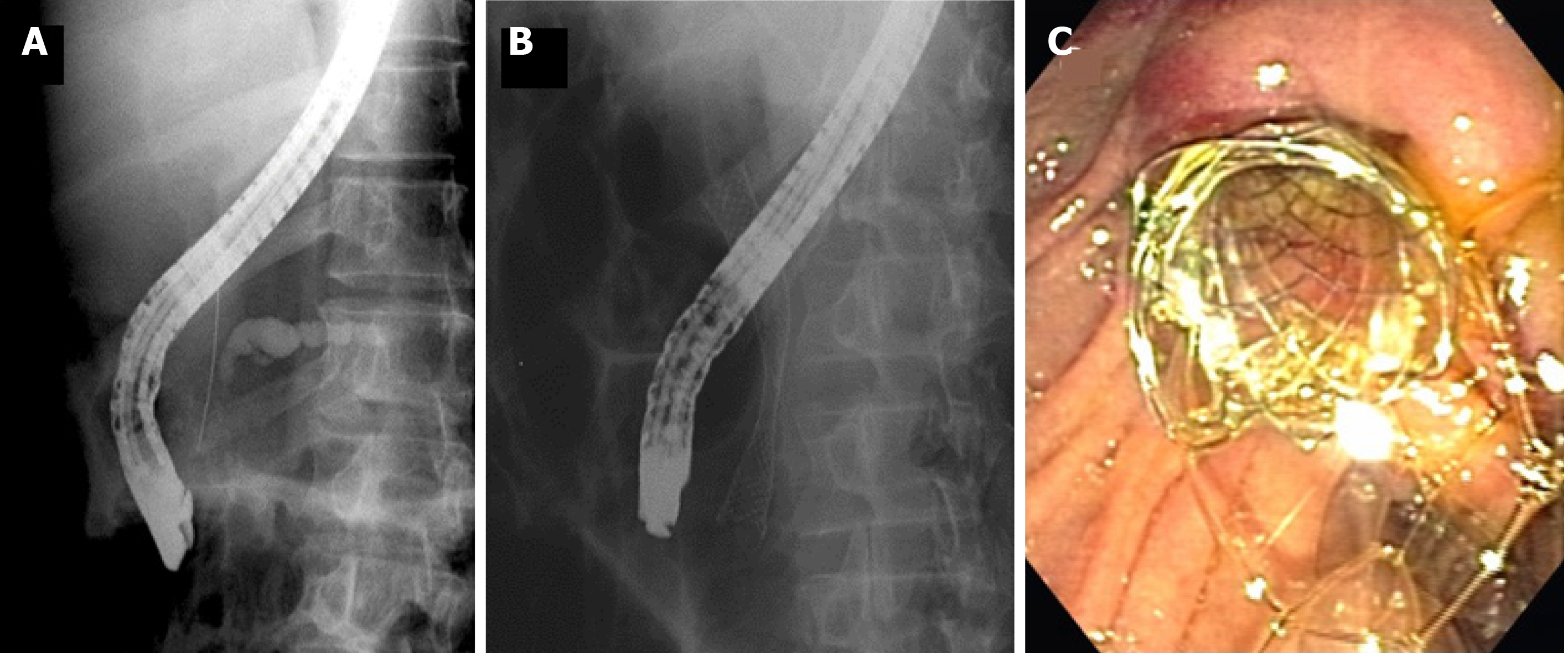
Figure 4 Malignant pancreatic stricture causing upstream pancreatic duct dilation[21].
A: Note that the wire was advanced into the bile duct during endoscopic retrograde cholangiopancreatography to place a biliary stent for palliation of obstructive jaundice; B and C: Placement of a metallic biliary stent for palliation of obstructive jaundice in a patient with unresectable pancreatic cancer [fluoroscopic picture (B); endoscopic picture (C)]. Citation: Porta M, Fabregat X, Malats N, Guarner L, Carrato A, de Miguel A, Ruiz L, Jariod M, Costafreda S, Coll S, Alguacil J, Corominas JM, Solà R, Salas A, Real FX. Exocrine pancreatic cancer: symptoms at presentation and their relation to tumour site and stage. Clin Transl Oncol 2005; 7: 189-197. Copyright© The Authors 2005. Published by Springer Nature.
- Citation: Zeeshan MS, Ramzan Z. Current controversies and advances in the management of pancreatic adenocarcinoma. World J Gastrointest Oncol 2021; 13(6): 472-494
- URL: https://www.wjgnet.com/1948-5204/full/v13/i6/472.htm
- DOI: https://dx.doi.org/10.4251/wjgo.v13.i6.472


