Copyright
©The Author(s) 2020.
World J Gastrointest Oncol. Aug 15, 2020; 12(8): 893-902
Published online Aug 15, 2020. doi: 10.4251/wjgo.v12.i8.893
Published online Aug 15, 2020. doi: 10.4251/wjgo.v12.i8.893
Figure 1 Representative rectal neuroendocrine tumor morphology and histochemistry.
A: a. Gross morphology of a G1 rectal neuroendocrine tumor (RNET) (black arrow heads the tumor); b. Gross morphology of a G2 RNET (black arrow heads the tumor); c. Gross morphology of a neuroendocrine carcinoma (black arrow heads the tumor). The diameter of the rectal tumor was approximately 2.0 cm, and it was 11 cm away from the anus. Pathological examination confirmed that it was a neuroendocrine carcinoma. B: a. Hematoxylin and eosin (HE) staining of a G1 RNET (× 100); b. HE staining of a G2 RNET (× 100); c. HE staining of a rectal neuroendocrine carcinoma (× 100); d. Ki-67 staining in a G1 RNET (black arrow heads the positive staining; × 100); e. Ki-67 staining in a G2 RNET (black arrow heads the positive staining; × 100); f. Ki-67 staining in an rectal neuroendocrine carcinoma (black arrow heads the positive staining; × 100).
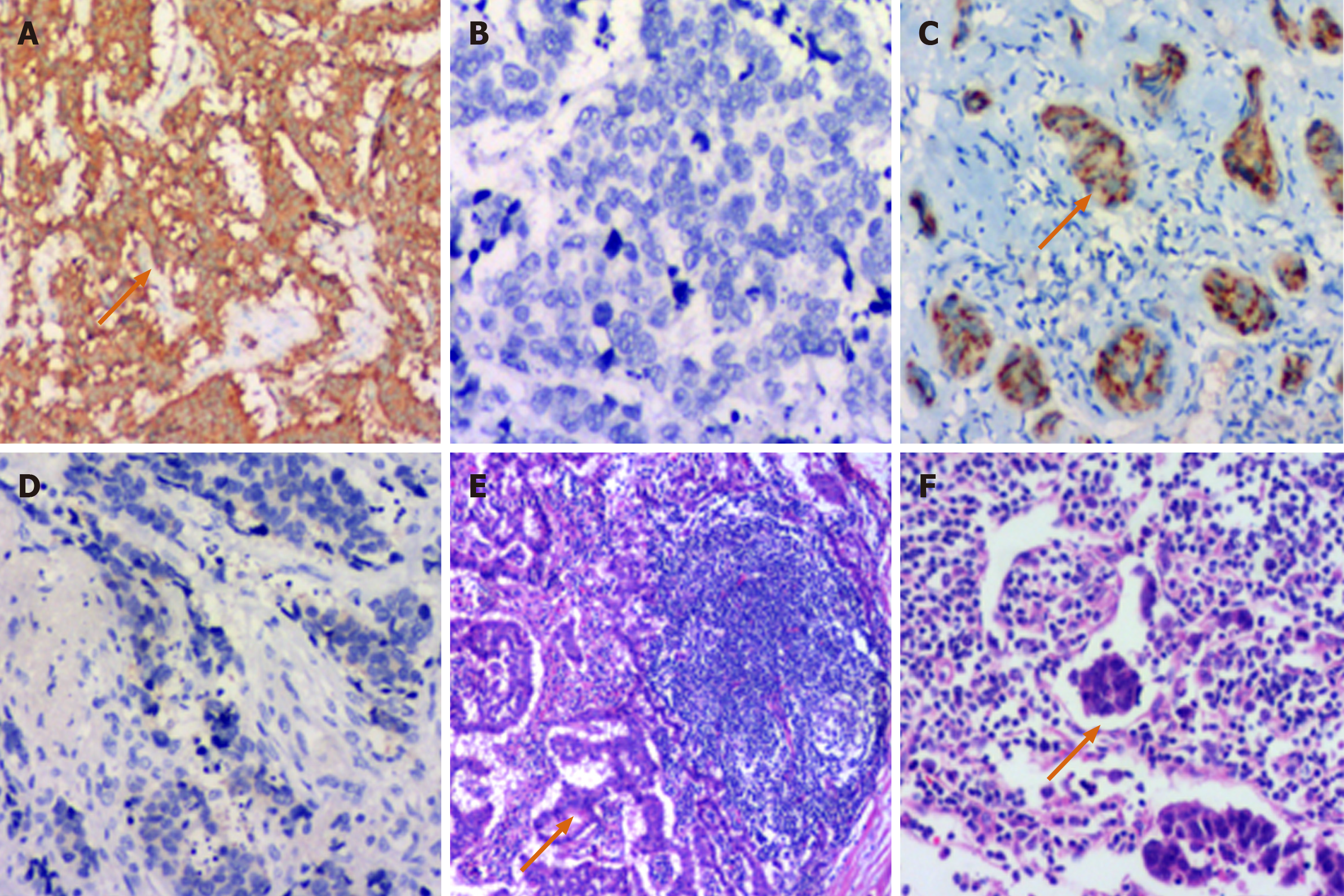
Figure 2 Immunohistochemical features.
A: Positive synaptophysin immunohistochemical staining in a G2 rectal neuroendocrine tumor (RNET) (black arrow heads the positive staining; × 100). B: Negative synaptophysin immunohistochemical staining in an RNEC (× 100); C: Positive chromogranin A immunohistochemical staining in a G1 RNET (black arrow heads the positive staining; × 100); D: Negative chromogranin A immunohistochemical staining in an RNEC (× 100); E: Lymph node metastasis of a G2 RNET (black arrow shows the tumor tissue; hematoxylin and eosin staining, × 100); F: Black arrow shows a thrombus in the lymph vessel (hematoxylin and eosin staining, × 100).
- Citation: Yu YJ, Li YW, Shi Y, Zhang Z, Zheng MY, Zhang SW. Clinical and pathological characteristics and prognosis of 132 cases of rectal neuroendocrine tumors. World J Gastrointest Oncol 2020; 12(8): 893-902
- URL: https://www.wjgnet.com/1948-5204/full/v12/i8/893.htm
- DOI: https://dx.doi.org/10.4251/wjgo.v12.i8.893


