Copyright
©The Author(s) 2023.
World J Stem Cells. Sep 26, 2023; 15(9): 947-959
Published online Sep 26, 2023. doi: 10.4252/wjsc.v15.i9.947
Published online Sep 26, 2023. doi: 10.4252/wjsc.v15.i9.947
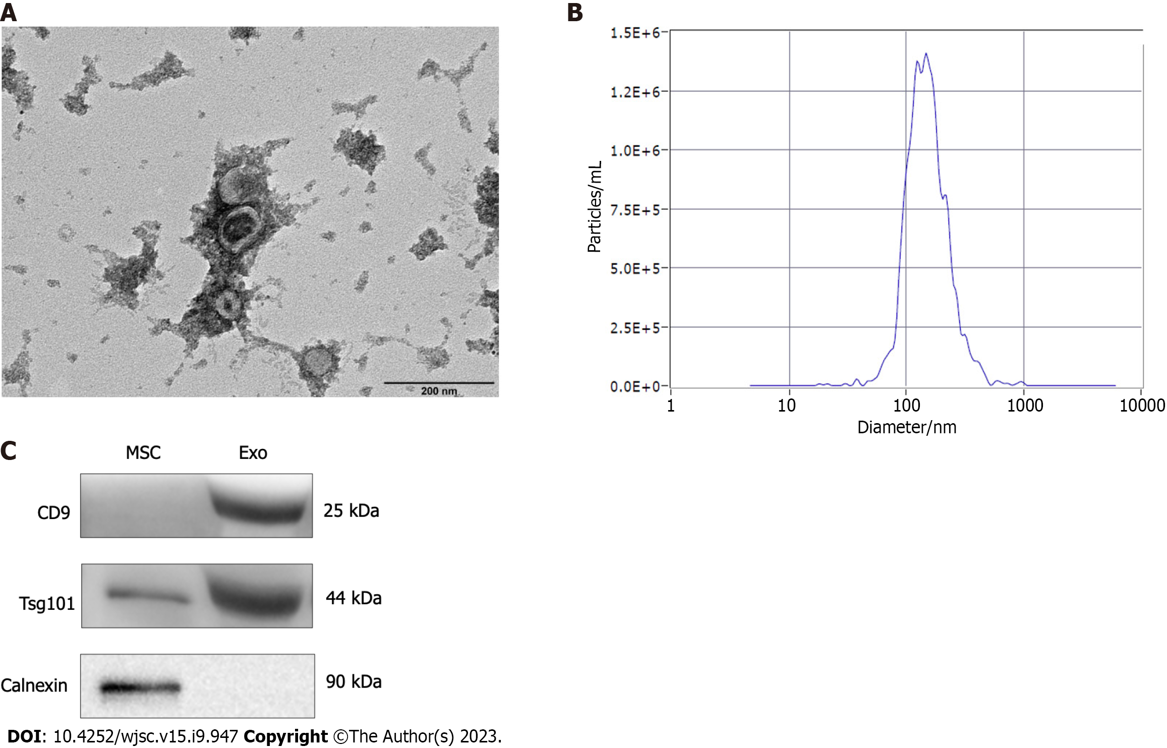
Figure 1 Characterization of human umbilical cord mesenchymal stem cell-derived exosomes.
A: Morphology of the exosomes assessed by transmission electron microscopy. Scale bar = 200 nm; B: Grain size of the exosomes; C: Surface protein markers of exosomes. MSC: Mesenchymal stem cell; Exo: Exosome.
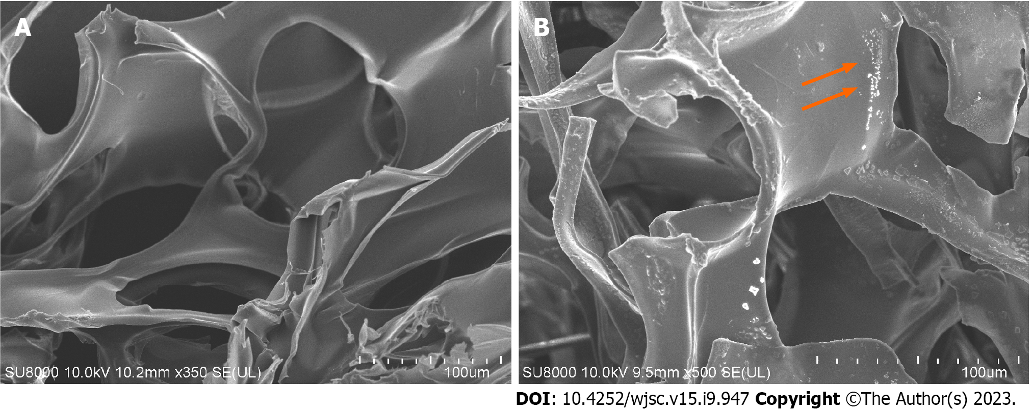
Figure 2 Characterization of gelatin sponge loaded with exosomes.
A: SEM image of the gelatin sponge; B: SEM images of the gelatin sponge loaded with exosomes. The orange arrows show the exosomes.
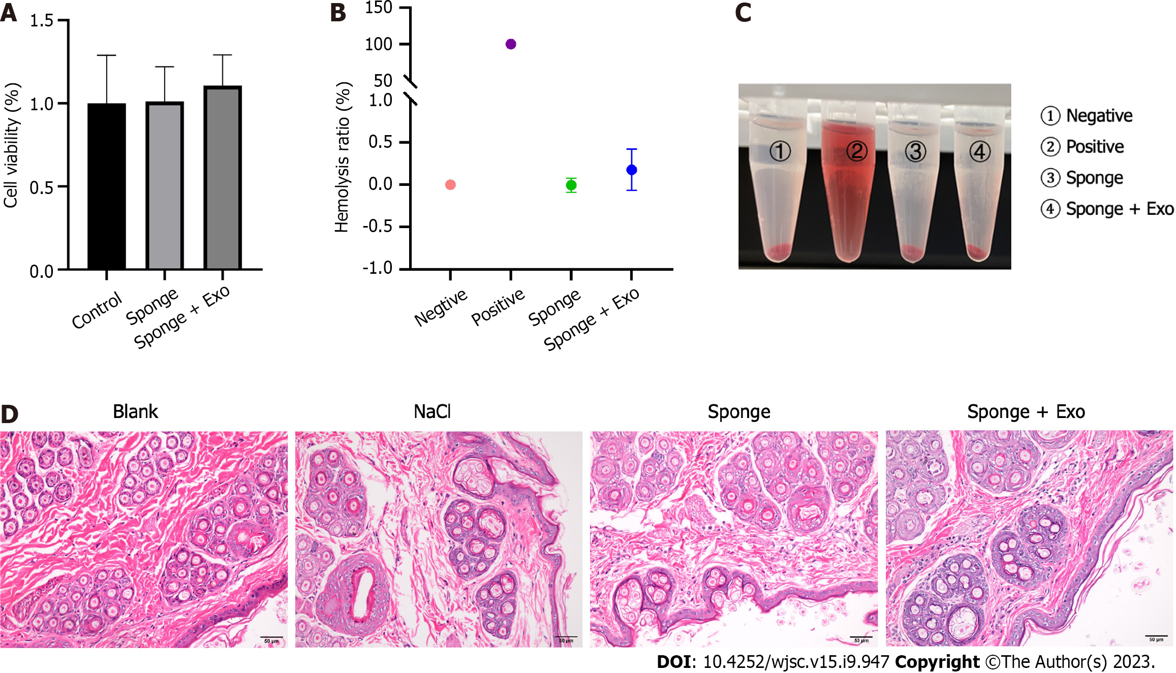
Figure 3 Biosafety of gelatin sponge loaded with exosomes.
A: Cell viability of gelatin sponge loaded with exosomes; B and C: Hemolysis ratio of gelatin sponge loaded with exosomes; D: Representative diagram of pathological condition of rabbit skin after stimulation with exosome and sponge extract. Scale bar = 50 μm. Exo: Exosome.
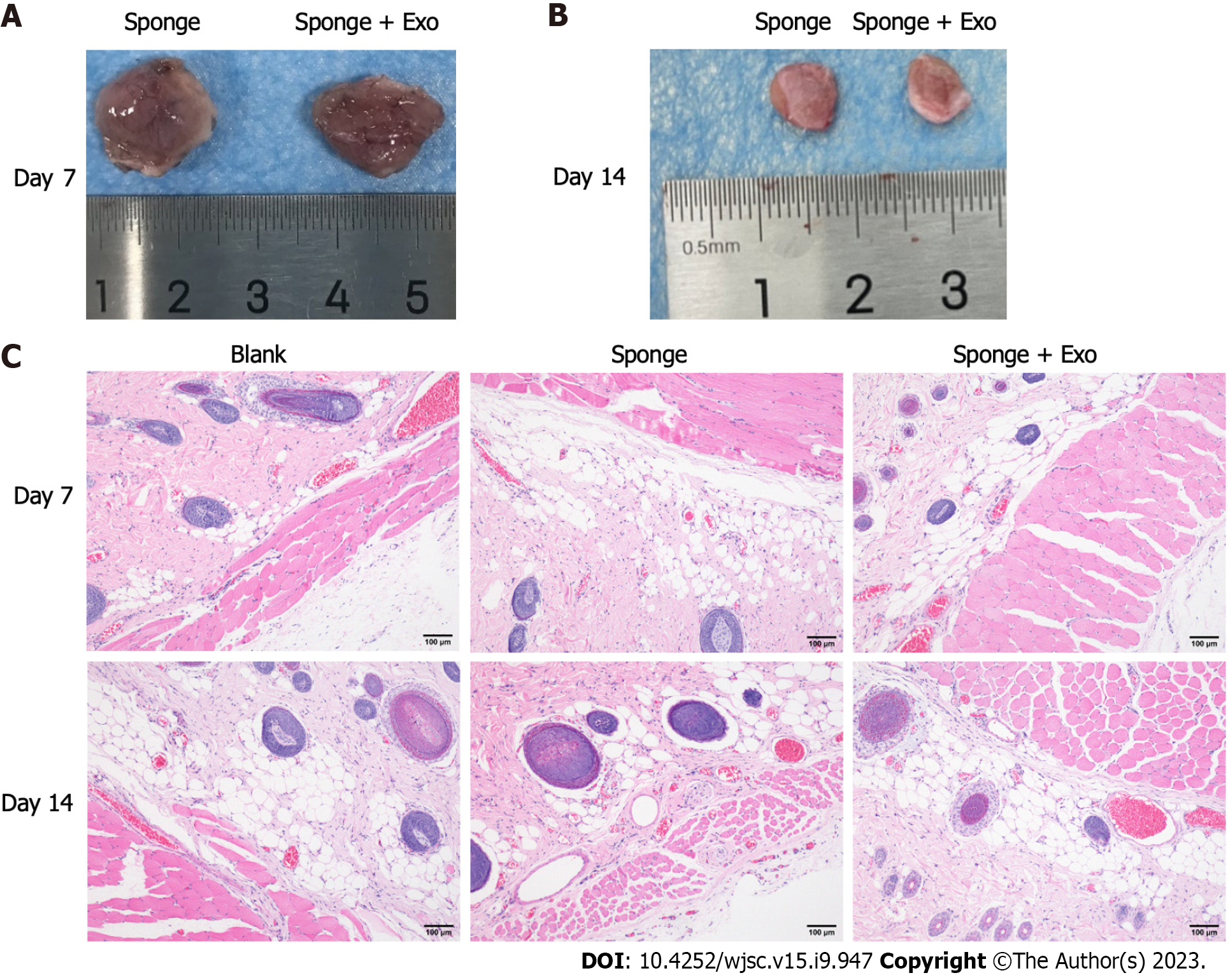
Figure 4 Biocompatibility of gelatin sponge loaded with exosomes.
A and B: The size of the gelatin sponge remaining on day 7 and day 14; C: Hematoxylin and eosin staining of the adjacent skin tissues in the dorsal subcutaneous space of SD rats after 7 and 14 d of implantation. Scale bar = 100 μm. Exo: Exosome.
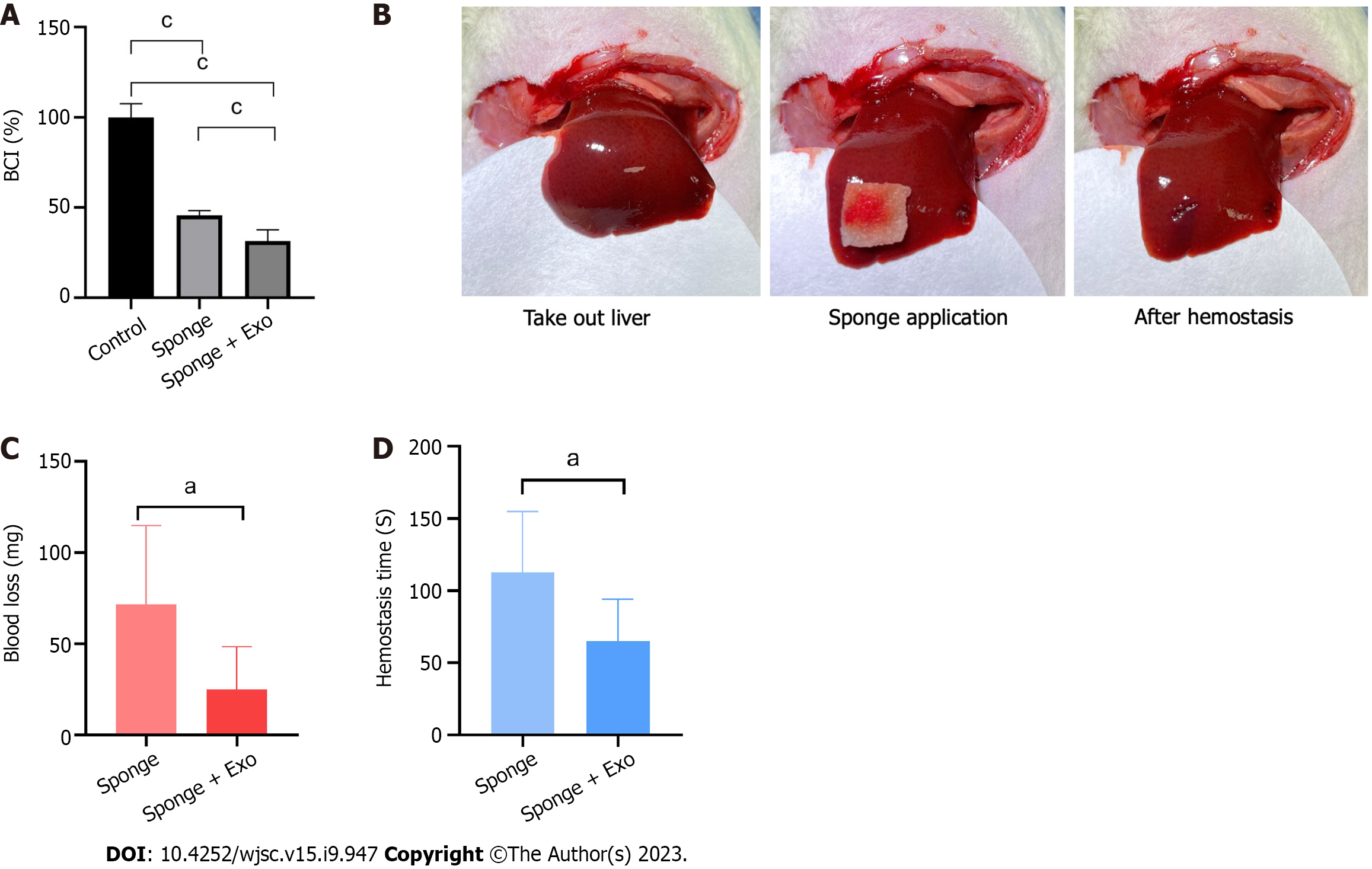
Figure 5 Hemostatic effect of gelatin sponge loaded with exosomes.
A: Clotting ability of exosome-loaded gelatin sponge; B: Experimental process of hemostasis in liver injury model; C: Hepatic blood loss in rats; D: Time required for hemostasis. aP < 0.05, cP < 0.001. n = 6. Exo: Exosome.
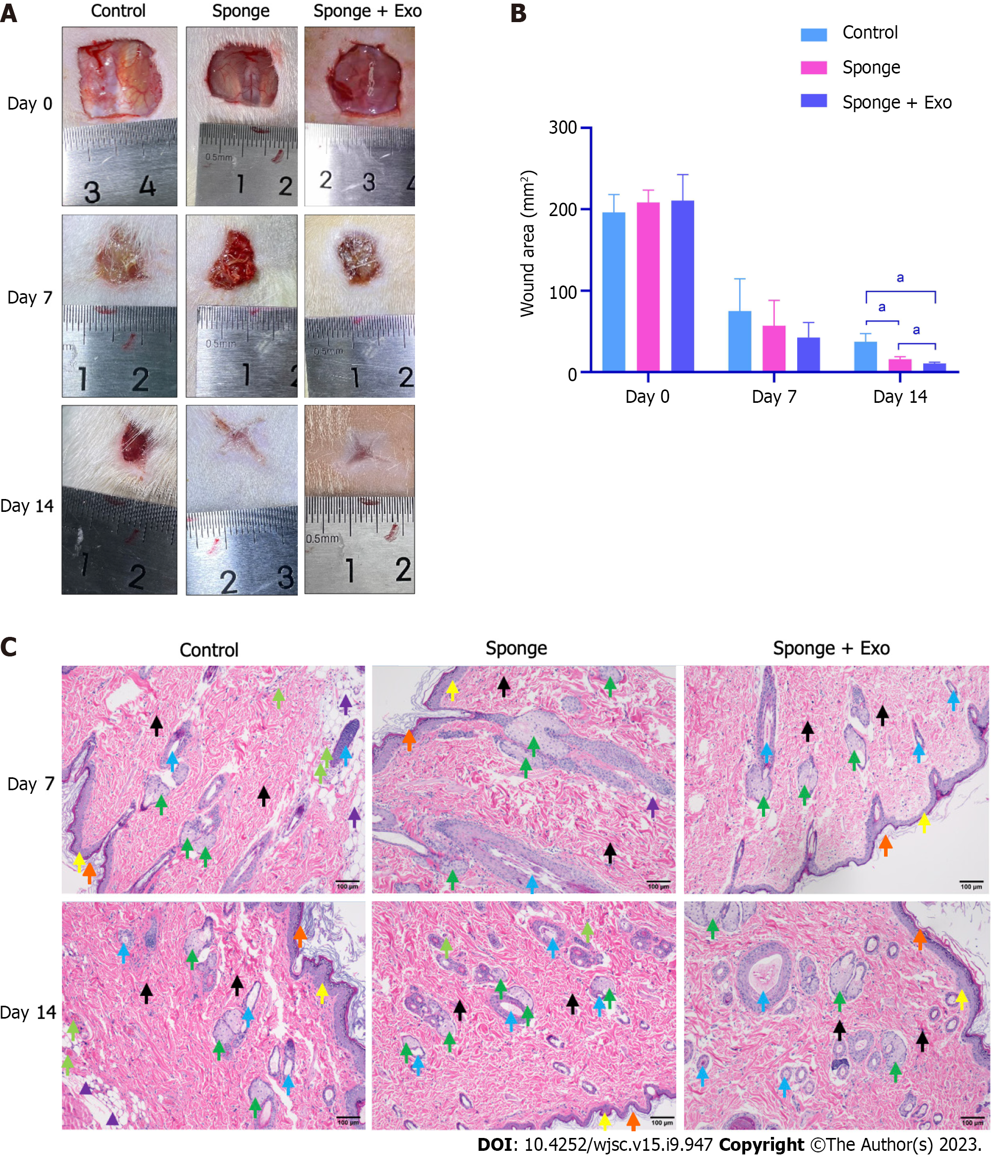
Figure 6 Wound healing effect of gelatin sponge loaded with exosomes.
A: Representative images of rat wounds; B: Statistical analysis of the wound area; C: Hematoxylin and eosin-stained wound tissue sections as observed under a light microscope. The orange arrows indicate the stratum corneum, the yellow arrows indicate the granular layer, the green arrows indicate the sebaceous gland, the blue arrows indicate the hair follicle, the black arrows indicate the collagen fibers, the light green arrows indicate the blood vessels, and the purple arrows indicate the subcutaneous fat. Scale bar = 100 μm. aP < 0.05. n = 3. Exo: Exosome.
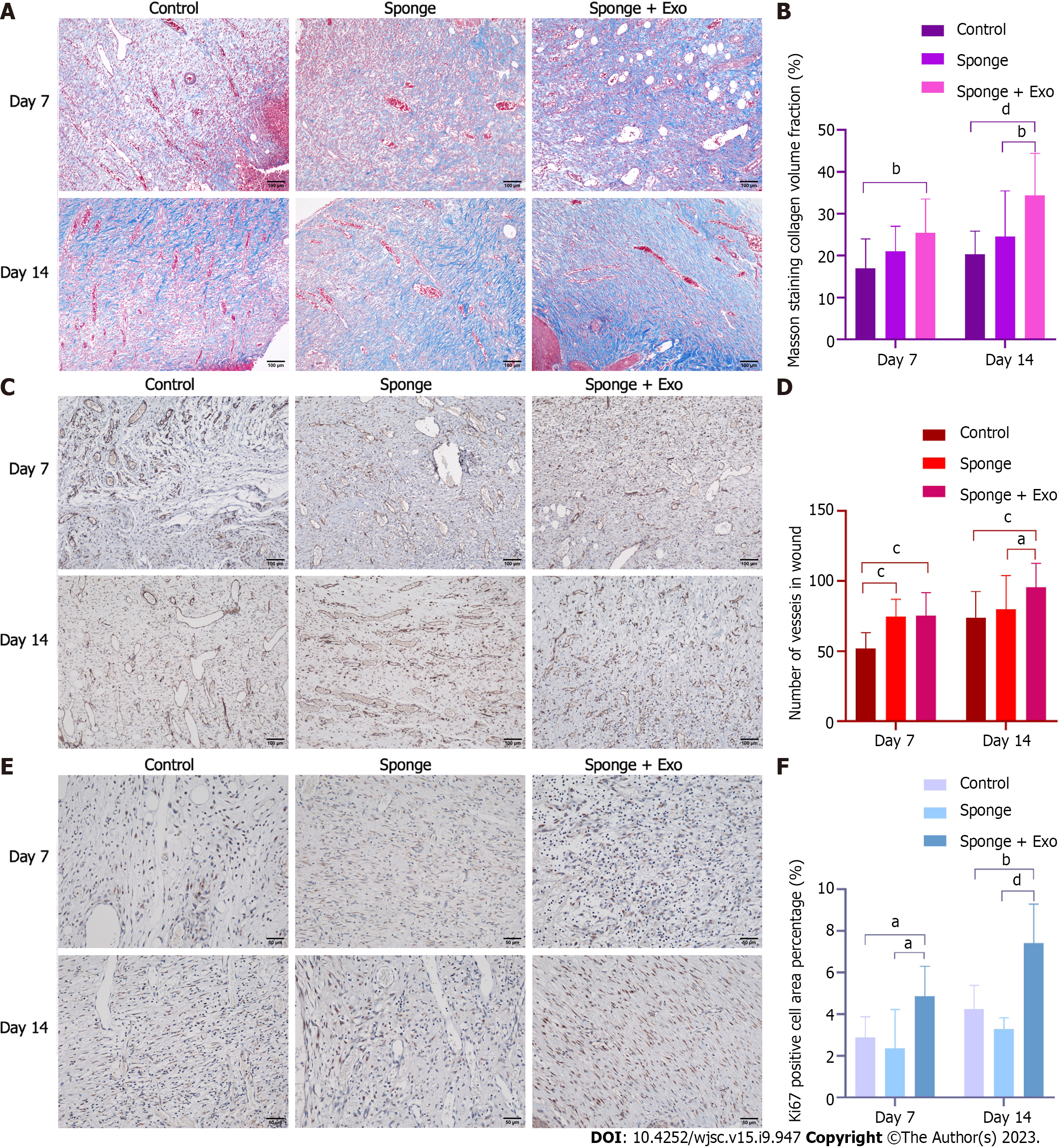
Figure 7 Gelatin sponge loaded with exosomes promotes collagen formation, cell proliferation, and angiogenesis during the wound healing process.
A: Masson-stained wound tissue sections as observed. Scale bar = 100 μm; B: Statistical analysis of the percentage of collagen volume with respect to tissue volume; C: CD31-positive vessels in wound tissue sections. Scale bar = 100 μm; D: Statistical analysis of CD31-positive vessels; E: Ki67-positive cells in wound tissue sections. Scale bar = 50 μm; F: Statistical analysis of Ki67 positive cell area percentage. aP < 0.05, bP < 0.01, cP < 0.001, dP < 0.0001. Exo: Exosome.
- Citation: Hu XM, Wang CC, Xiao Y, Jiang P, Liu Y, Qi ZQ. Enhanced wound healing and hemostasis with exosome-loaded gelatin sponges from human umbilical cord mesenchymal stem cells. World J Stem Cells 2023; 15(9): 947-959
- URL: https://www.wjgnet.com/1948-0210/full/v15/i9/947.htm
- DOI: https://dx.doi.org/10.4252/wjsc.v15.i9.947


