Copyright
©The Author(s) 2022.
World J Gastroenterol. Apr 28, 2022; 28(16): 1656-1670
Published online Apr 28, 2022. doi: 10.3748/wjg.v28.i16.1656
Published online Apr 28, 2022. doi: 10.3748/wjg.v28.i16.1656
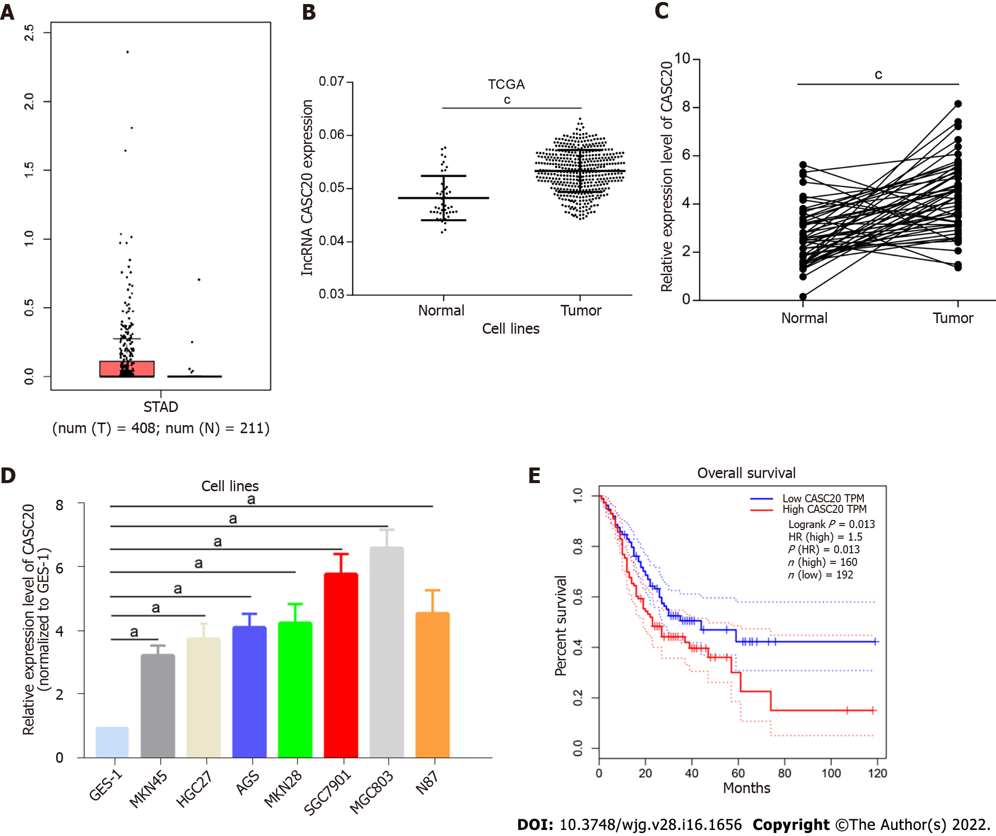
Figure 1 High level of cancer susceptibility 20 in gastric cancer tissues and cell lines.
A and B: Cancer susceptibility 20 (CASC20) expression in gastric cancer (GC) samples and normal samples from TCGA GC database; C: CASC20 expression in 50 pairs of clinically collected gastric cancer tissues and normal tissue samples; D: CASC20 expression in GC cell lines and GES-1 cell line was analyzed by quantitative real-time polymerase chain reaction; E: Overall survival curves of TCGA GC patients with different CASC20 expression. aP < 0.05 and cP < 0.001 vs control.
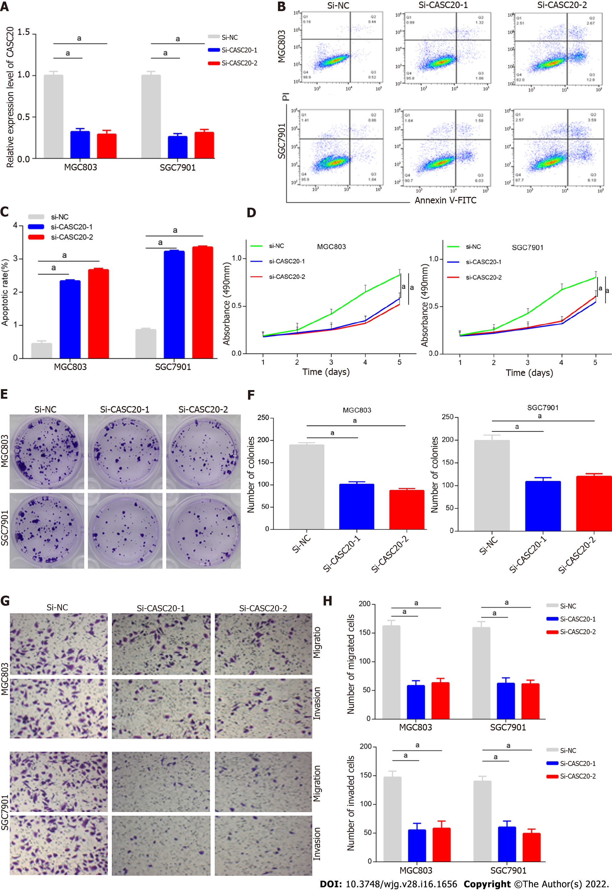
Figure 2 Konckdown of cancer susceptibility 20 inhibits the apoptosis, proliferation and metastasis of gastric cancer cells.
A: Efficacy assessment of cancer susceptibility 20 (CASC20) silence through quantitative real-time polymerase chain reaction method in MGC803 and SGC7901 cells; B and C: Cell apoptosis evaluation in transfected cells through flow cytometry; D: Cell viability was verified by CCK-8 experiments after transfection with si-CASC20; E and F: Colony formation assay was used to detect the cell proliferation ability after transfection with si-CASC20; G and H: Transwell assay was employed for examining the effect of CASC20 low-expression on gastric cancer cell migration and invasion. aP < 0.05 vs control.
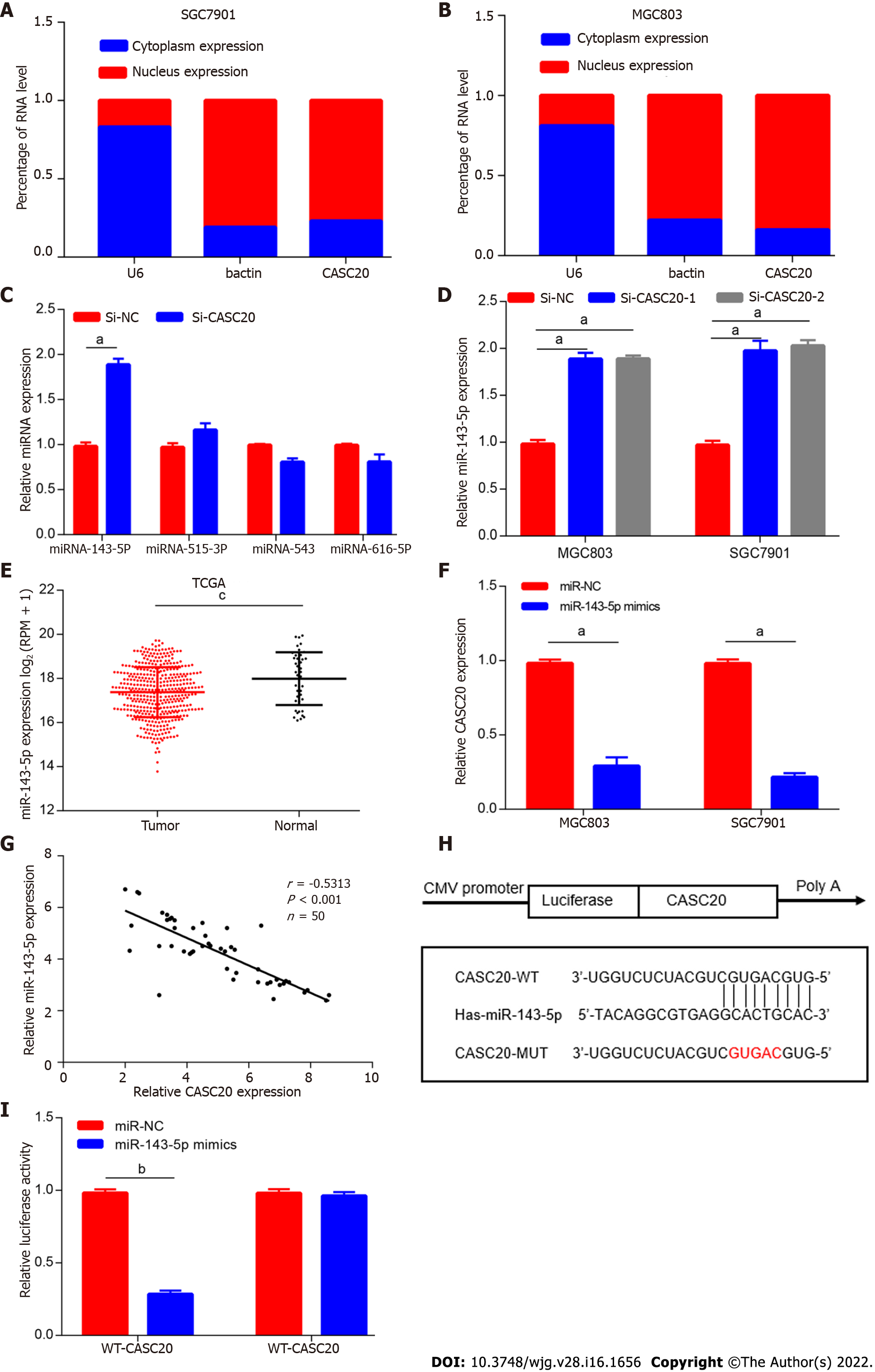
Figure 3 miR-143-5p served as a target of cancer susceptibility 20 in gastric cancer cells.
A and B: Relative cancer susceptibility 20 (CASC20) expression levels in nucleus and cytoplasm of SGC7901 and MGC803 cells; C: The relative expressions of miRNAs in gastric cancer (GC) cells after downregulation of CASC20; D: The expression of miR-143-5p was detected by quantitative real-time polymerase chain reaction in GC cells after transfection with si-CASC20; E: The expression levels of miR-143-5p in GC from TCGA database; F: The detection of CASC20 expression after transfection of miR-143-5p mimics; G: The correlation analysis between miR-143-5p expression and CASC20 expression in 50 GC tissues; H: Bioinformatics presentation of CASC20 and miR-143-5p binding sites; I: The verification of CASC20 and miR-143-5p interaction by luciferase report assay. aP < 0.05, bP < 0.01 and cP < 0.001 vs control.
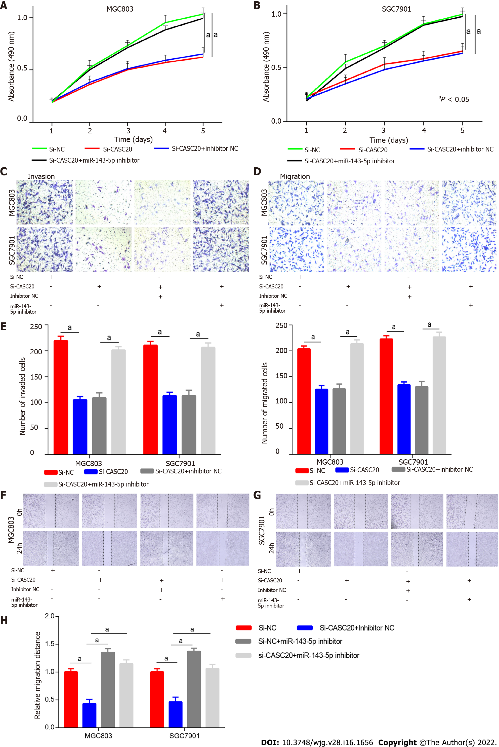
Figure 4 The regulation of cancer susceptibility 20 on gastric cancer cells is mediated by miR-143-5p.
A and B: Gastric cancer (GC) cells were treated with si-NC, si-cancer susceptibility 20 (CASC20), si-CASC20 + inhibitor NC, si-CASC20 + miR-143-5p inhibitor, CCK-8 assays evaluated the proliferation; C-E: Transwell assays tested invasion and migration abilities of transfected cells; F-H: GC cells were treated with si-NC, si-CASC20 + inhibitor NC, si-NC + miR-143-5p inhibitor, si-CASC20 + miR-143-5p inhibitor, wound healing assay was used to detect migration ability. aP < 0.05 vs control.
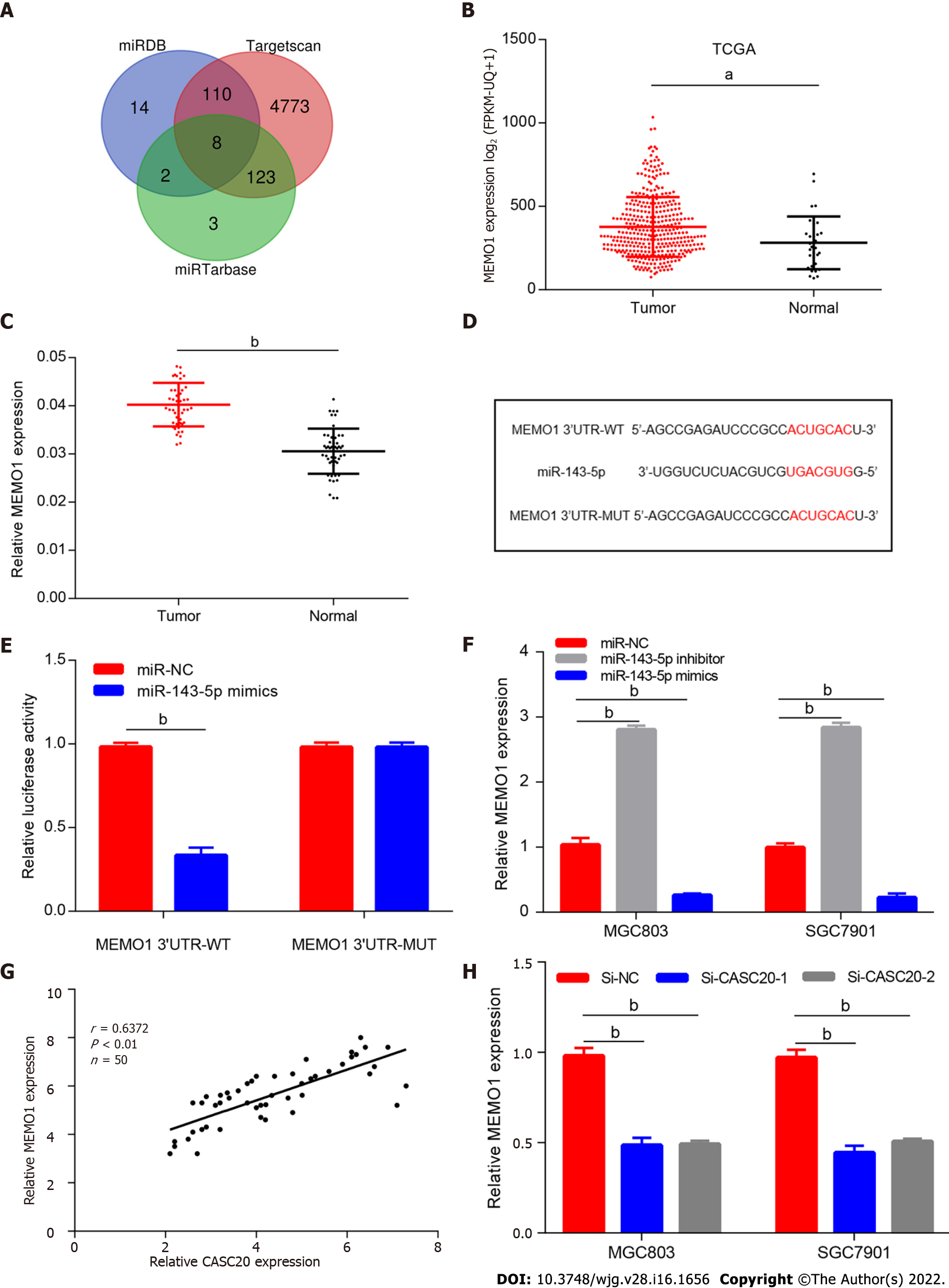
Figure 5 Cancer susceptibility 20 regulates the miR-143-5p target MEMO1.
A: Bioinformatic tools were used to predict the potential target mRNA of miR-143-5p; B: The expression levels of MEMO1 in gastric cancer (GC) from TCGA STAD database; C: MEMO1 expression in 50 pairs of gastric cancer tissues and normal tissue samples; D: Putative miR-143-5p binding sequence and mutation sequence of MEMO1 mRNA were as shown; E: Dual luciferase reporter assays were used to confirm the direct target between miR-143-5p and MEMO1; F: MEMO1 expression was decreased in GC cells transfected by miR-143-5p mimics; G: The correlation analysis between cancer susceptibility 20 (CASC20) and MEMO1 expression in 50 GC tissues; H: MEMO1 expression was decreased after knockdown of CASC20. aP < 0.05, bP < 0.01 vs control.
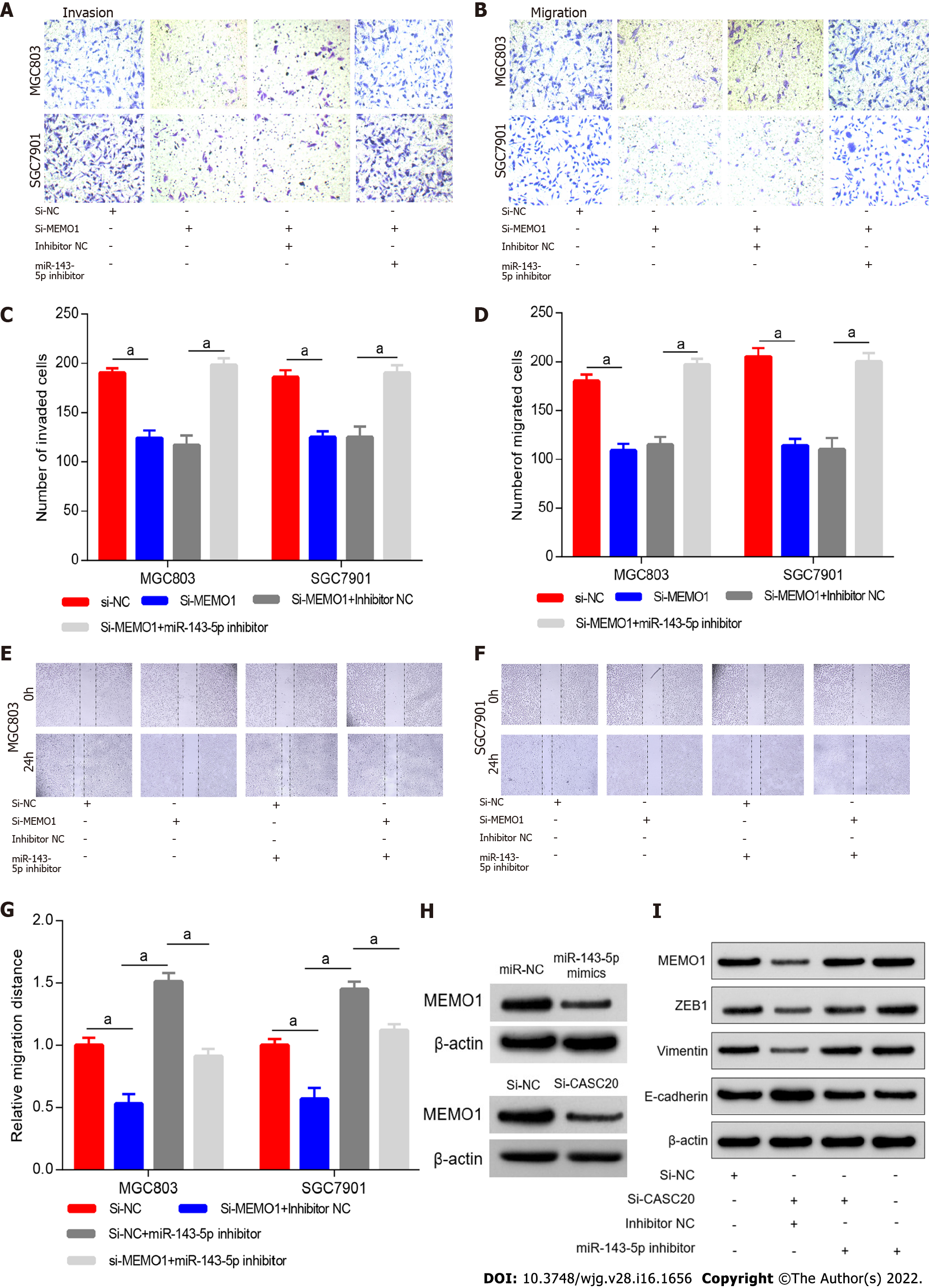
Figure 6 Inhibition of miR-143-5p blocked the function of MEMO1 knockdown in gastric cancer cells.
A-D: MGC803 and SGC7901 cells were transfected with si-NC, si-MEMO1, si-MEMO1 + inhibitor NC, si-MEMO1 + miR-143-5p inhibitor, respectively. Transwell assay was employed for the detection of cell invasion and migration ability; E-G: MGC803 and SGC7901 cells were transfected with si-NC, si-MEMO1 + inhibitor NC, si-NC + miR-143-5p inhibitor, si-MEMO1 + miR-143-5p inhibitor, respectively. Wound healing assay was used to detect migration ability; H: The protein expression of MEMO1 was detected by western blot assay after transfecting with miR-143-5p mimics or si-cancer susceptibility 20; I: Western blot was utilized to detect the expressions of EMT-related proteins (ZEB1, Vimentin and E-cadherin). aP < 0.05 vs control.
- Citation: Shan KS, Li WW, Ren W, Kong S, Peng LP, Zhuo HQ, Tian SB. LncRNA cancer susceptibility 20 regulates the metastasis of human gastric cancer cells via the miR-143-5p/MEMO1 molecular axis. World J Gastroenterol 2022; 28(16): 1656-1670
- URL: https://www.wjgnet.com/1007-9327/full/v28/i16/1656.htm
- DOI: https://dx.doi.org/10.3748/wjg.v28.i16.1656


