Published online Nov 26, 2021. doi: 10.12998/wjcc.v9.i33.10286
Peer-review started: May 27, 2021
First decision: July 15, 2021
Revised: July 15, 2021
Accepted: October 14, 2021
Article in press: October 14, 2021
Published online: November 26, 2021
Processing time: 179 Days and 3.8 Hours
Stapedial tendon ossification is a rare disease, with only a few reports. The stapedial tendon originates from the apex of the pyramidal eminence and is attached to the neck of the stapes. In stapedial tendon ossification, the stapes is fixed, causing conductive hearing loss. In most cases, complete hearing rest
A 28-year-old woman presented to our hospital with the major complaint of bilateral hearing loss that started during childhood. Exploratory tympanotomy was performed due to suspicion of otosclerosis or middle ear anomalies. We found bilateral conductive hearing loss due to stapedial tendon ossification with a middle ear anomaly during surgery. There have been several reports of complete recovery of hearing after resection of the stapedial tendon. However, in this case, recovery of hearing was insufficient, even with the division of the stapedial tendon. In the second surgery, the stapes anomaly and footplate fixation were confirmed, and hearing was completely recovered after stapedotomy. Therefore, we report this case with a review of the relevant literature.
This is the first case of stapedial tendon ossification and fixation of the footplate surgically diagnosed on both sides. With surgical treatment, successful results are expected.
Core Tip: To date, only a few papers have reported stapedial tendon ossification. Hearing is completely restored by dividing the stapedial tendon. However, as in our case, when hearing improvement after surgery is insufficient, the possibility of accompanying malformations should be suspected, and secondary surgery should be considered. Successful results can be obtained when the comorbid anomaly is resolved.
- Citation: Yoo JS, Lee CM, Yang YN, Lee EJ. Complete restoration of congenital conductive hearing loss by staged surgery: A case report. World J Clin Cases 2021; 9(33): 10286-10292
- URL: https://www.wjgnet.com/2307-8960/full/v9/i33/10286.htm
- DOI: https://dx.doi.org/10.12998/wjcc.v9.i33.10286
Stapedial ankylosis is a congenital or an acquired ossification of the stapes, which causes conductive hearing loss through disturbance of the sound transmission system. The main cause is fixation of the footplate along with the normal superstructure, such as otosclerosis, tympanic sclerosis, and congenital stapes fixation. However, since 1957, cases with limited movement of the stapes due to ossification of the stapedial tendon have been reported[1-14]. The stapedial tendon originates from the apex of the pyramidal eminence, attaches to the neck of the stapes, and is involved in the acoustic reflex. Due to this anatomical location, stapedial tendon ossification was also referred to as the elongation of the pyramidal eminence or bony bar[2-4]. When the stapes is fixed, this causes conductive hearing loss. In most cases, complete hearing im
Bilateral hearing loss.
A 28-year-old woman visited our clinic with bilateral hearing loss. She had poor hearing since childhood.
There was no previous history of infection or trauma, and there were no other medical symptoms.
She had no family history of hearing loss.
On otoscopy, both eardrums were normal.
On the patient’s first visit to our clinic, laboratory test results were within the normal ranges.
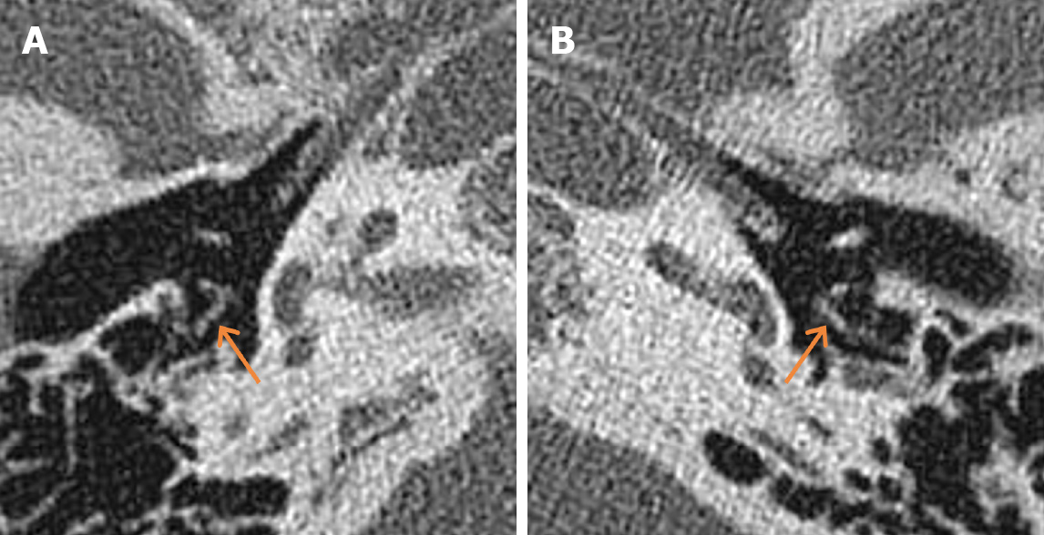
Temporal bone CT showed normal middle ear and ossicular chain.
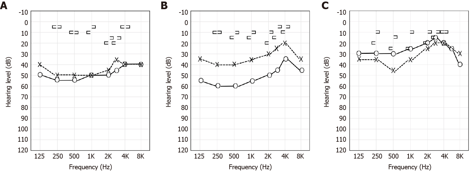
As per pure tone audiometry, the air-conduction threshold was 50 dB on the right and 45 dB on the left, and bilateral conductive hearing loss was found, showing a 40 dB air-bone gap (Figure 1A). The speech discrimination scores on both sides were 92% each.
The patient was diagnosed with stapedial tendon ossification and fixation of the footplate on both sides.
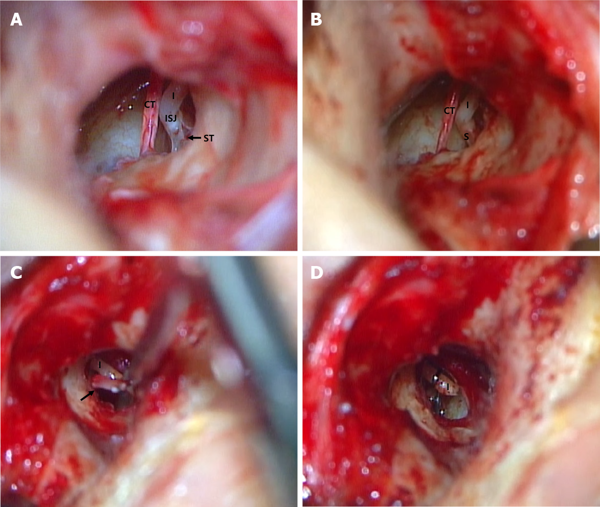
Right-sided stapes surgery was planned due to suspicion of bilateral otosclerosis. When we scanned the temporal bone CT before surgery, the bilateral stapedial tendon showed a slightly thicker appearance than normal, but was overlooked (Figure 2). An exploratory tympanotomy was performed on the right side under general anesthesia. Entire fixed ossicle mobility was also identified. After separating the incudostapedial joint (I-S joint), it was found that the stapedial tendon was ossified. It looked like a bony bar between the pyramidal eminence and the stapes neck. The posterior crura and footplates of the stapes were invisible and appeared in the form of a monopod. After division of the stapedial tendon with a fine burr, we noted that the movement of the stapes became hypermobile, and that it was misdiagnosed as otosclerosis. The separated I-S joint was connected with fibrin glue, and the surgery was terminated. The hearing test after surgery showed that the air-bone gap (ABG) on the right side decreased by 5 dB. One month later, an exploratory tympanotomy was performed on the contralateral side, and the same stapedial tendon ossification was observed; only the stapedial tendon was divided with a fine burr without any other treatment (Figure 3). After dividing only the stapedial tendon on the left side, the mobility of the stapes improved. Because the patient was under general anesthesia, we ended the operation in anticipation of proper hearing recovery. The hearing test after surgery showed that the ABG on the left side decreased by approximately 15 dB (Figure 1B). Even if the right side was separated from the I-S joint incorrectly and the improvement was insufficient, the ABG on the left side improved by only 15 dB; therefore, we considered the possibility of an accompanying abnormality and planned a second surgery on the right side. During the second surgery, the stapes were observed in the form of a rod, and it was found that the footplate was fixed. Therefore, stapedotomy was planned. After measuring the length between the incus and the stapes, a small hole was made with a perforator, and a window was made on the posterior side of the footplate using a Skeeter drill. A piston wire (0.4 mm × 5 mm) was hung over the long process of the incus and fixed using a crimper. The operation was subsequently terminated.
There were no postoperative complications. One year after surgery, her hearing in the right ear had markedly improved (Figure 1C). Therefore, we are considering a left-sided stapedotomy and expecting the same improvement; however, due to the coronavirus disease 2019, it is planned in the next few years.
The major cause of isolated stapedial ankylosis is stapedial footplate fixation with normal stapes suprastructure due to otosclerosis, congenital stapedial fixation, and tympanosclerosis as reported by Henner[15]. In addition to stapedial tendon ossification, there are various types of ankylosis caused by elongation of the pyramidal eminence, stapes promontory fixation, and stapes pyramidal bony bar with normal stapedial tendon. Ossification of the stapedius tendon rarely occurs due to congenital causes. If it occurs due to an acquired cause, such as infection, it can be accompanied by tympanosclerosis. This disease, which needs to be differentiated, is otosclerosis and is often misdiagnosed. Stapedial tendon ossification has been reported in only a few studies[1-14]. Most previous reports have shown bilateral conductive hearing loss without trauma or past history. Here, on otoscopy, the eardrum was almost normal, and the patient did not complain of any other symptoms. From previous reports showing the same findings over different generations, a congenital cause was found, and it was thought to have an autosomal recessive inheritance[4,9].
Stapedial tendon ossification can be diagnosed using preoperative CT and exploratory tympanotomy. On temporal bone CT, it can be diagnosed as a linear image from the pyramidal eminence to the stapes superstructure[7]. Based on these findings, it can be differentiated from otosclerosis[16]. A definitive diagnosis is to directly confirm ossification of the stapedius tendon that fixes the stapes through tympanotomy. The treatment involves dividing the tendon between the stapes and the pyramidal eminence. After securing a sufficient surgical field, it is carefully performed using a microdrill or laser. Hearing was significantly restored following division of the stapedial tendon. The average ABG before surgery was 30 dB, which closed after the surgery[2-6,10] (Table 1).
| Ref. | Ears | Diagnosis | Treatment | Average of air-bone gaps | ||
| [pre OP/post OP/△ (pre OP-post OP)] | ||||||
| Schuknecht et al[1], 1957 | 1 | PE elongation | Division | NA | NA | NA |
| Patel[5], 1972 | 1 | ST ossification | Division and stapedectomy | 51.67 dB | 15 dB | 36.67 dB |
| Cremers et al[2], 1986 | 2 | PE elongation | Division | 22.5 dB | 0 dB | 22.5 dB |
| Grant et al[6], 1991 | 4 | ST ossification | Division | 31.67 dB | 2.5 dB | 29.17 dB |
| Jecker et al[8], 1992 | 2 | ST ossification | Division | NA | NA | NA |
| Kinsella et al[3], 1993 | 3 | Bony bar | Division | 29.44 dB | 0 dB | 29.44 dB |
| Kurosaki et al[7], 1995 | 8 | ST ossification | Division | NA | NA | NA |
| 1 | ST ossification | Division and stapedectomy | NA | NA | NA | |
| Thies et al[4], 1996 | 5 | Bony bar | Division | 25.83 dB | NA | NA |
| 3 | ST ossification | Division | NA | NA | NA | |
| Hara et al[9], 1997 | 8 | ST ossification | Division | NA | NA | NA |
| Lee et al[11], 2009 | 1 | ST ossification | Division | 30 dB | 13.33 dB | 16.67 dB |
| Wetmore et al[12], 2011 | 6 | Bony bar | Division | NA | NA | NA |
| Ulkü[13], 2011 | 2 | ST ossification | Division | NA | NA | NA |
| Wasano et al[10], 2014 | 2 | ST ossification | Division | 25 dB | 2.5 dB | 22.5 dB |
| Chan[14], 2016 | 1 | Bony bar | Division | NA | NA | NA |
Our patient had no history of ear infection or degenerative change in the middle ear; thus, the stapedial tendon ossification might have originated from a congenital lesion. Our patient was misdiagnosed with bilateral otosclerosis, and at the time of the first right side surgery, we made a mistake in separating the right I-S joint. However, on the left side, even though only the stapedial tendon was divided because the lesion was the same, hearing improvement after surgery was insufficient. Therefore, we suspected an accompanying malformation and performed the second surgery, wherein we found an anomaly of the stapes in the form of a monorod and fixation of the footplate. Thus, a window was made on the posterior side of the fixed footplate using a Skeeter drill, and a piston wire (4 mm × 0.5 mm) was hung on the incus long process and inserted into the window. Hearing on the right side showed marked improvement after the stapedotomy. Previously, one case of stapedial tendon ossification and fixation of the footplate on both sides was reported. At that time, only one side was diagnosed and treated with stapedectomy[7]. This is relevant because our case is the first to have been surgically diagnosed on both sides. In addition, successful results are expected after surgical treatment.
Congenital stapedial footplate fixation (CSFF) is a rare cause of congenital conductive hearing loss. Nevertheless, this is a common congenital ossicle abno
Stapedial tendon ossification is a very rare disease that results in bilateral conductive hearing loss. If stapedial tendon ossification is found, good progress can be expected following division. In most previously reported case reports, the ABG was completely restored after surgery. However, as in our case, when hearing improvement after surgery is insufficient, the possibility of accompanying malformations should be considered. Thus, secondary surgery may be necessary, and successful results can be obtained when the comorbid anomaly is resolved.
Provenance and peer review: Unsolicited article; Externally peer reviewed.
Specialty type: Otorhinolaryngology
Country/Territory of origin: South Korea
Peer-review report’s scientific quality classification
Grade A (Excellent): 0
Grade B (Very good): B
Grade C (Good): 0
Grade D (Fair): D
Grade E (Poor): 0
P-Reviewer: you Y, Zhou S S-Editor: Wu YXJ L-Editor: Webster JR P-Editor: Wu YXJ
| 1. | Schuknecht HF, Trupiano S. Some interesting middle ear problems. Laryngoscope. 1957;67:395-409. [RCA] [PubMed] [DOI] [Full Text] [Cited by in Crossref: 26] [Cited by in RCA: 26] [Article Influence: 0.4] [Reference Citation Analysis (0)] |
| 2. | Cremers CW, Hoogland GA. Congenital stapes ankylosis by elongation of the pyramidal eminence. Communication. Ann Otol Rhinol Laryngol. 1986;95:167-168. [RCA] [PubMed] [DOI] [Full Text] [Cited by in Crossref: 14] [Cited by in RCA: 14] [Article Influence: 0.4] [Reference Citation Analysis (0)] |
| 3. | Kinsella JB, Kerr AG. Familial stapes superstructure fixation. J Laryngol Otol. 1993;107:36-38. [RCA] [PubMed] [DOI] [Full Text] [Cited by in Crossref: 15] [Cited by in RCA: 15] [Article Influence: 0.5] [Reference Citation Analysis (0)] |
| 4. | Thies C, Handrock M, Sperling K, Rcis A. Possible autosomal recessive inheritance of progressive hearing loss with stapes fixation. J Med Genet. 1996;33:597-599. [RCA] [PubMed] [DOI] [Full Text] [Cited by in Crossref: 7] [Cited by in RCA: 7] [Article Influence: 0.2] [Reference Citation Analysis (0)] |
| 5. | Patel KN. Ossification of the stapedius tendon. J Laryngol Otol. 1972;86:863-865. [RCA] [PubMed] [DOI] [Full Text] [Cited by in Crossref: 13] [Cited by in RCA: 12] [Article Influence: 0.2] [Reference Citation Analysis (0)] |
| 6. | Grant WE, Grant WJ. Stapedius tendon ossification: a rare cause of congenital conductive hearing loss. J Laryngol Otol. 1991;105:763-764. [RCA] [PubMed] [DOI] [Full Text] [Cited by in Crossref: 13] [Cited by in RCA: 13] [Article Influence: 0.4] [Reference Citation Analysis (0)] |
| 7. | Kurosaki Y, Kuramoto K, Matsumoto K, Itai Y, Hara A, Kusakari J. Congenital ossification of the stapedius tendon: diagnosis with CT. Radiology. 1995;195:711-714. [RCA] [PubMed] [DOI] [Full Text] [Cited by in Crossref: 11] [Cited by in RCA: 11] [Article Influence: 0.4] [Reference Citation Analysis (0)] |
| 8. | Jecker P, Hartwein J. [Ossification of the stapedius tendon as a rare cause of conductive hearing loss]. Laryngorhinootologie. 1992;71:344-346. [RCA] [PubMed] [DOI] [Full Text] [Cited by in Crossref: 4] [Cited by in RCA: 5] [Article Influence: 0.2] [Reference Citation Analysis (0)] |
| 9. | Hara A, Ase Y, Kusakari J, Kurosaki Y. Dominant hereditary conductive hearing loss due to an ossified stapedius tendon. Arch Otolaryngol Head Neck Surg. 1997;123:1133-1135. [RCA] [PubMed] [DOI] [Full Text] [Cited by in Crossref: 8] [Cited by in RCA: 8] [Article Influence: 0.3] [Reference Citation Analysis (0)] |
| 10. | Wasano K, Kanzaki S, Ogawa K. Bilateral congenital conductive hearing loss due to ossification of the stapedius tendon. Otol Neurotol. 2014;35:e119-e120. [RCA] [PubMed] [DOI] [Full Text] [Cited by in Crossref: 4] [Cited by in RCA: 4] [Article Influence: 0.4] [Reference Citation Analysis (0)] |
| 11. | Lee KS, Park KH, Lee CK. A Case of Unilateral Conductive Hearing Loss Treated with Stapedial Tenotomy. Korean J Otorhinolaryngol-Head Neck Surg. 2009;52:449-452. [RCA] [DOI] [Full Text] [Cited by in Crossref: 2] [Cited by in RCA: 2] [Article Influence: 0.1] [Reference Citation Analysis (0)] |
| 12. | Wetmore SJ, Gross AF. Congenital fixation of the head of the stapes in three family members. Ear Nose Throat J. 2011;90:360-366. [RCA] [PubMed] [DOI] [Full Text] [Cited by in Crossref: 5] [Cited by in RCA: 5] [Article Influence: 0.4] [Reference Citation Analysis (0)] |
| 13. | Ulkü CH. [Isolated stapedius tendon ossification: a case report]. Kulak Burun Bogaz Ihtis Derg. 2011;21:95-97. [RCA] [PubMed] [DOI] [Full Text] [Cited by in Crossref: 2] [Cited by in RCA: 2] [Article Influence: 0.1] [Reference Citation Analysis (0)] |
| 14. | Chan KC. Stapes-pyramidal fixation by a bony bar. Ear Nose Throat J. 2016;95:E40-E41. [RCA] [PubMed] [DOI] [Full Text] [Cited by in Crossref: 3] [Cited by in RCA: 3] [Article Influence: 0.4] [Reference Citation Analysis (0)] |
| 15. | Henner R. Congenital middle ear malformations. AMA Arch Otolaryngol. 1960;71:454-458. [RCA] [PubMed] [DOI] [Full Text] [Cited by in Crossref: 41] [Cited by in RCA: 41] [Article Influence: 0.6] [Reference Citation Analysis (0)] |
| 16. | Kösling S, Plontke SK, Bartel S. Imaging of otosclerosis. Rofo. 2020;192:745-753. [RCA] [PubMed] [DOI] [Full Text] [Cited by in Crossref: 2] [Cited by in RCA: 8] [Article Influence: 1.6] [Reference Citation Analysis (0)] |
| 17. | Tolisano AM, Fontenot MR, Nassiri AM, Hunter JB, Kutz JW Jr, Rivas A, Isaacson B. Pediatric Stapes Surgery: Hearing and Surgical Outcomes in Endoscopic vs Microscopic Approaches. Otolaryngol Head Neck Surg. 2019;161:150-156. [RCA] [PubMed] [DOI] [Full Text] [Cited by in Crossref: 13] [Cited by in RCA: 13] [Article Influence: 2.2] [Reference Citation Analysis (0)] |
| 18. | Totten DJ, Marinelli JP, Carlson ML. Incidence of Congenital Stapes Footplate Fixation Since 1970: A Population-based Study. Otol Neurotol. 2020;41:489-493. [RCA] [PubMed] [DOI] [Full Text] [Cited by in Crossref: 3] [Cited by in RCA: 4] [Article Influence: 1.0] [Reference Citation Analysis (0)] |


