Published online Oct 26, 2021. doi: 10.12998/wjcc.v9.i30.9129
Peer-review started: June 3, 2021
First decision: June 25, 2021
Revised: June 26, 2021
Accepted: August 18, 2021
Article in press: August 18, 2021
Published online: October 26, 2021
Processing time: 139 Days and 18 Hours
Foreign body in the deep neck is mostly associated with accidental ingestion of the animal spiculate bone which penetrates the full-thickness of the aerodigestive tract into the fascial spaces of the neck. In general, perforation of the esophagus often results in periesophagitis and even fatal abscesses. The presence of a giant foreign body in the neck without obvious symptoms or complications for many years is rare.
We present the case of a 32-year-old man who intentionally swallowed a thermometer which was unable to be located by endoscopy at his initial visit. He had no remarkable symptoms for 5 years other than paresthesia and limited movement of the left neck until 7 d before this admission. The foreign body was removed successfully by the surgery.
The presence of a giant foreign body in the neck without obvious symptoms or complications for many years is rare. Both endoscopic and radiological examinations are essential for the diagnosis of alimentary foreign bodies.
Core Tip: Foreign body in the deep neck is mostly associated with accidental ingestion of the animal spiculate bone. These emergency accidents need timely diagnosis and treatment, otherwise they might cause fatal complications such as neck abscess or macrovascular injury. We present the case of a 32-year-old man who intentionally swallowed a thermometer which was unable to be located by endoscopy at his initial visit. He had no remarkable symptoms for 5 years as the long-term retention of the foreign body resulted in the formation of the surrounding fibrous membrane. This case implies that both endoscopic and radiological examinations are essential for the diagnosis of alimentary foreign bodies.
- Citation: Yang L, Li W. Unusual cervical foreign body - a neglected thermometer for 5 years: A case report. World J Clin Cases 2021; 9(30): 9129-9133
- URL: https://www.wjgnet.com/2307-8960/full/v9/i30/9129.htm
- DOI: https://dx.doi.org/10.12998/wjcc.v9.i30.9129
Most of the foreign bodies deep in the neck are related to the accidental swallowing of sharp bones of animals. Timely diagnosis and treatment are critical, otherwise neck abscess, bleeding, and other fatal complications could not be avoided. The presence of a foreign body deep in the neck without obvious history, symptoms, signs, and complications for many years is extremely rare. Here we report a patient who intentionally swallowed a mercury thermometer, which was embedded in the neck without obvious symptoms for 5 years. This case implies that both endoscopic and radiological examinations are essential for the diagnosis of alimentary foreign bodies. A foreign body entrapped by thick fibrous tissue can be ignored in an asymptomatic carrier, resulting in long-term retention.
A 32-year-old man with a history of drug abuse presented to the emergency department with deterioration of paresthesia and limited movement of the left neck for 7 d.
The patient had slight paresthesia of the neck for 5 years. Seven days before this admission, the symptom became intolerable and was accompanied by a limited movement of the neck after an acute upper respiratory tract infection. He perceived a foreign body existing on the left side of the neck and the feeling was severe while coughing or swallowing.
The patient had no history of neck trauma, cervical spondylosis, or contagious diseases.
The patient was a heroin abuser 5 years ago and achieved successful detoxification for 2 years. He had no family history of hereditary diseases.
There was no abnormality in vital signs. Physical examination revealed only mild pain during the palpation of the left neck, and a rod-shaped solid substance was touched. Its location was parallel with the long axis of the left cervical artery.
A blood test revealed a slightly elevated C-reactive protein level (1.53 mg/dL) and white blood cell count (10.94 × 10⁹/L). Other blood test results were normal.
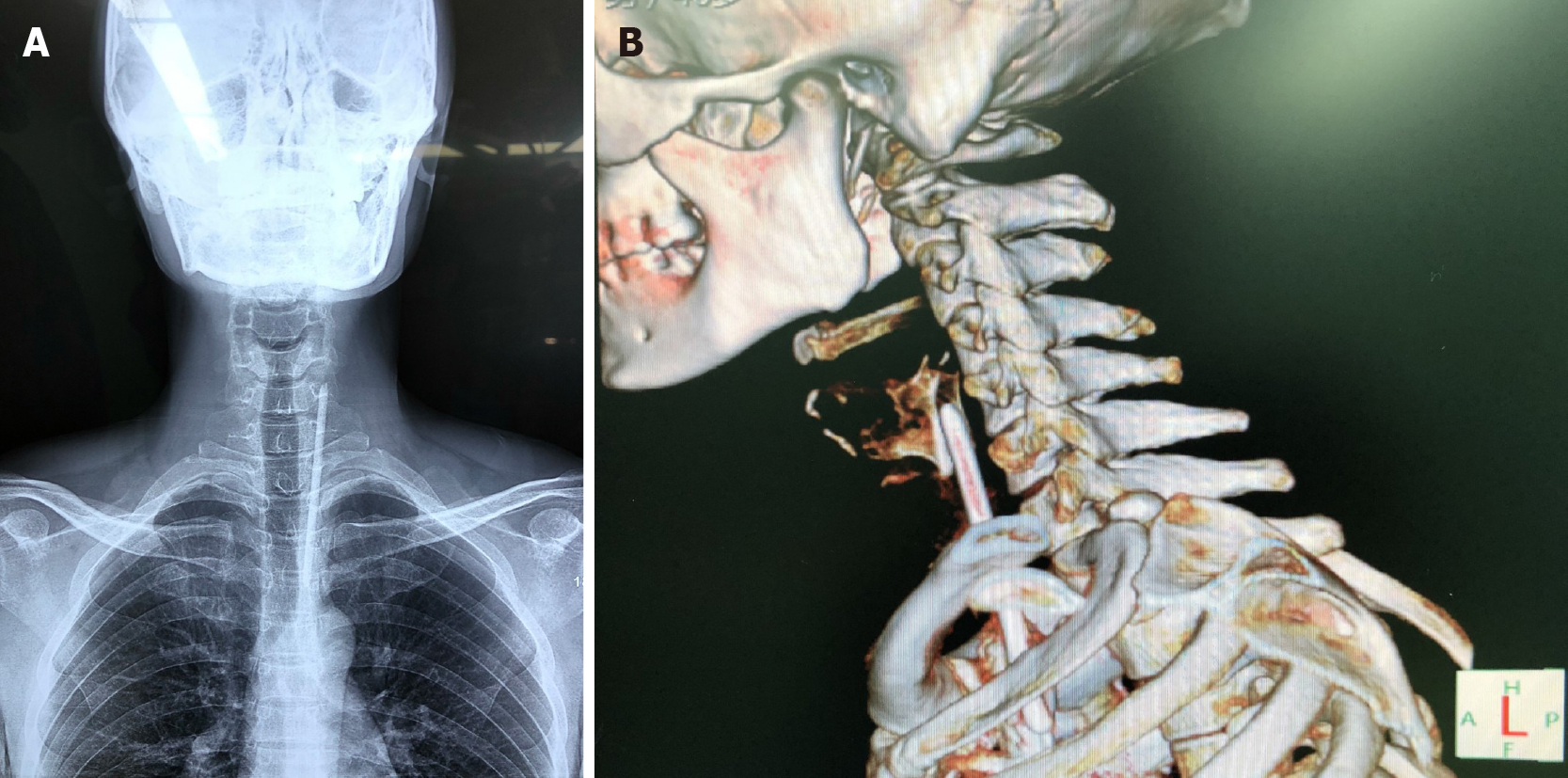
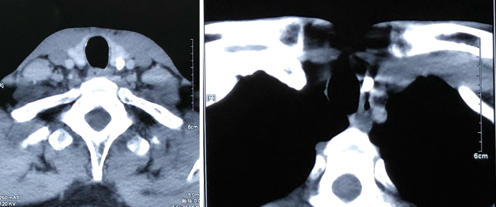
The radiological examination showed that a rod-shaped foreign body was diagonally located in the anterior region of the neck and the lower end of the rod extended to the thorax (Figure 1) and computed tomography revealed a high density extending from the cervical region to the upper mediastinal area (Figure 2).
Because of the unexpected imaging findings, further medical history was acquired. He finally admitted that he deliberately swallowed a segment of mercury thermometer after he broke a mercury thermometer and poured out the mercury 5 years ago under the influence of drugs when he was alone. He was sent to the emergency department immediately to undergo an endoscopy after he told his family about the accident. But neither a foreign body nor trauma or perforation was found. He did not receive further examinations or treatments because the clinician thought that he was talking nonsense under the influence of drugs, and the patient’s symptoms were tolerable until 7 d before this admission.
The final diagnosis was esophageal perforation caused by a cervical foreign body.
The patient was admitted to the Otolaryngology and Head & Neck Surgery Department for surgery. Lateral neck incision for foreign body exploration was proposed, and performed after informed consent was obtained from the patient.
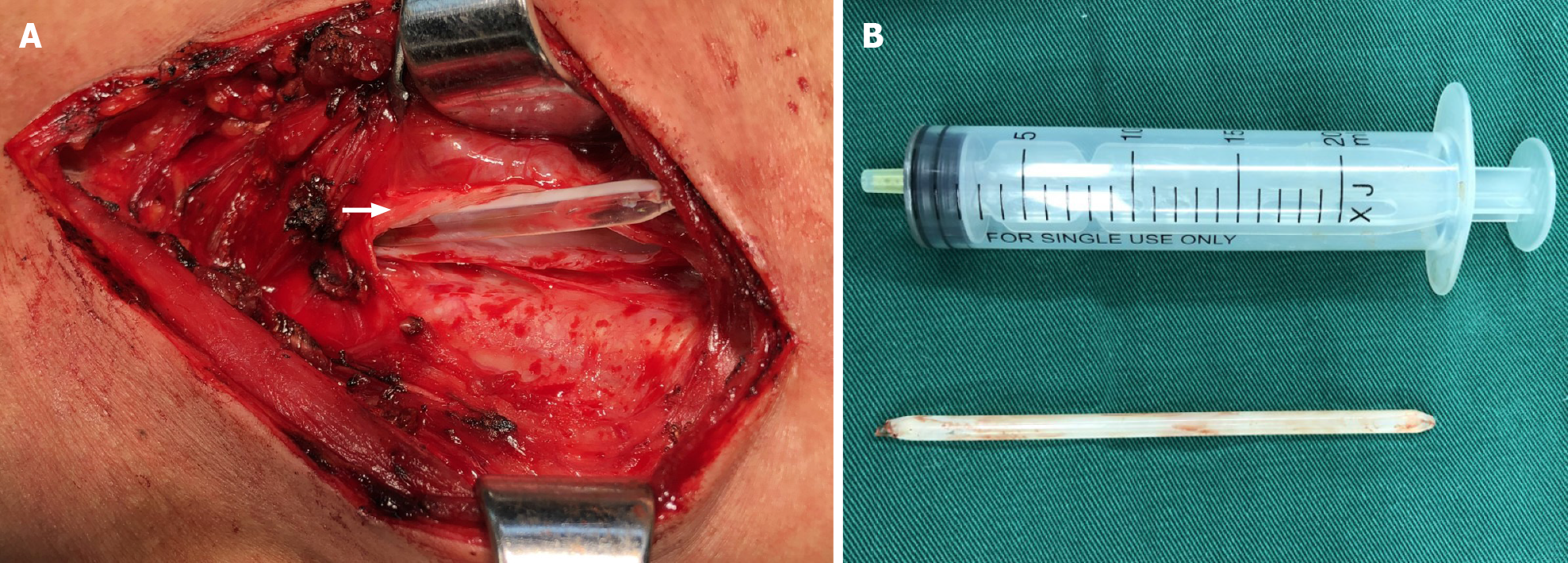
The surgical field displayed that the thermometer was outside the esophageal wall and was located in the left tracheoesophageal groove and wrapped in the fibrous membrane. The foreign body was removed successfully (Figure 3). The incision primarily healed and he was discharged after 5 d with no complications.
Metallic or plastic foreign bodies of small size can persist in the deep neck without infection and symptoms for a long time[1,2]. In general, perforation of the esophagus often results in periesophagitis and even fatal abscesses[3]. But it is not the case for this patient, in whom the thermometer could be detected because of symptoms and signs after his self-mutilation swallowing. It was supposed that the residual mercury of the broken thermometer played the role of disinfection and promoted esophageal perforation healing[4]. In addition, the glass material and smooth surface of the thermometer also facilitated the fibrous envelop formation. This resulted in a lack of symptoms and delay in diagnosis and treatment. Furthermore, a radiological examination did not find the segment of the thermometer, which also constituted a flaw in the diagnostic procedure. Both this case and the previous similar cases imply that the slender and giant foreign body with spiculate end might penetrate the fascial spaces of the neck through the indiscoverable puncture hole[5].
A technical point to be emphasized is the management of the lower end of the thermometer. Frankly, it is hard to tell if the sharp tip of the thermometer has penetrated the wall of the aortic arch. This made massive hemorrhage possible during the removal of the thermometer. The dense fibrous wall around the thermometer made us believe that if there was bleeding, the dense fibrous capsule channel in the middle of the neck was the most likely way of drainage, which made the bleeding easy to control, because the lower end would also be welded to the aortic arch by dense fibrous tissue.
This case extended our knowledge about a sharp esophageal foreign body of big size. Emergency doctors should pay attention to the medical history of the patients and find the whereabouts of a foreign body. Both endoscopy and radiological examination are essential for the diagnosis of alimentary foreign bodies.
Manuscript source: Unsolicited manuscript
Specialty type: Surgery
Country/Territory of origin: China
Peer-review report’s scientific quality classification
Grade A (Excellent): A
Grade B (Very good): 0
Grade C (Good): C
Grade D (Fair): 0
Grade E (Poor): 0
P-Reviewer: Chen Y, Liakina V S-Editor: Yan JP L-Editor: Wang TQ P-Editor: Liu JH
| 1. | Li M, Xiang X, Li W, Lei D. [Foreign body in neck for 40 years with nodular goiter: 1 case report]. Zhonghua Er Bi Yan Hou Tou Jing Wai Ke Za Zhi. 2014;49:960. [RCA] [PubMed] [DOI] [Full Text] [Cited by in RCA: 1] [Reference Citation Analysis (0)] |
| 2. | Ozturk K, Turhal G, Gode S, Yavuzer A. Migration of a swallowed blunt foreign body to the neck. Case Rep Otolaryngol. 2014;2014:646785. [RCA] [PubMed] [DOI] [Full Text] [Full Text (PDF)] [Cited by in Crossref: 2] [Cited by in RCA: 4] [Article Influence: 0.4] [Reference Citation Analysis (0)] |
| 3. | Peng A, Li Y, Xiao Z, Wu W. Study of clinical treatment of esophageal foreign body-induced esophageal perforation with lethal complications. Eur Arch Otorhinolaryngol. 2012;269:2027-2036. [RCA] [PubMed] [DOI] [Full Text] [Cited by in Crossref: 39] [Cited by in RCA: 48] [Article Influence: 3.7] [Reference Citation Analysis (0)] |
| 4. | Batchu H, Chou HN, Rakowski D, Fan PL. The effect of disinfectants and line cleaners on the release of mercury from amalgam. J Am Dent Assoc. 2006;137:1419-1425. [RCA] [PubMed] [DOI] [Full Text] [Cited by in Crossref: 15] [Cited by in RCA: 11] [Article Influence: 0.6] [Reference Citation Analysis (0)] |
| 5. | Fine S, Watson JB, Habr F. Now you see it, endo you don't: case of the disappearing knife. Gastroenterology. 2013;144:e6-e7. [RCA] [PubMed] [DOI] [Full Text] [Cited by in Crossref: 1] [Cited by in RCA: 1] [Article Influence: 0.1] [Reference Citation Analysis (0)] |


