Published online Sep 16, 2021. doi: 10.12998/wjcc.v9.i26.7954
Peer-review started: May 17, 2021
First decision: June 15, 2021
Revised: June 17, 2021
Accepted: July 12, 2021
Article in press: July 12, 2021
Published online: September 16, 2021
Processing time: 115 Days and 17.1 Hours
Angiolipoma has been reported in many cases, and it often occurs in the skin of the trunk and limbs. However, angiolipoma in the scrotum is a rare disease with unknown etiology. This condition is difficult to diagnosis with other lumps in the scrotum.
A 32-year-old man presented to the urinary department with a history of an enlarged left scrotum with increasing discomfort for about 5 years. Physical examination revealed that there were a palpable mass measuring about 7.0 cm × 6.5 cm in the left scrotum, with smooth surfaces but without tenderness or adhesion to the skin. Ultrasound showed that there was a hyperechoic mass under the skin of the top scrotum, about 72 mm × 64 mm × 21 mm in size, with clear borders, uneven internal echo, and abundant blood flow signals. Serum human chorionic gonadotropin and alpha-fetoprotein were in normal level. Subcutaneous mass resection at the bottom of the left scrotum was performed under local anesthesia with 1% lidocaine. Postoperative pathological examination resulted in a diagnosis of subcutaneous angiolipoma of the scrotum. No evidence of recurrence was found at 6 mo after surgery and there were no complaints of discomfort.
Angiolipoma is an extremely rare type of benign tumor extremely rarely found in the scrotum, but needs to be considered when evaluating scrotal masses especially when the mass is solid. According to the characteristics of angiolipoma, surgical resection is the best treatment strategy and it is not prone to recurrence after resection.
Core Tip: Subcutaneous angiolipoma is a rare type of benign tumor extremely rarely found in the scrotum but needs to be considered when evaluating scrotal masses. We report a case of subcutaneous angiolipoma of the scrotum, and summarize its clinical manifestations and pathological characteristics to improve the understanding of this disease.
- Citation: Li SL, Zhang JW, Wu YQ, Lu KS, Zhu P, Wang XW. Subcutaneous angiolipoma in the scrotum: A case report. World J Clin Cases 2021; 9(26): 7954-7958
- URL: https://www.wjgnet.com/2307-8960/full/v9/i26/7954.htm
- DOI: https://dx.doi.org/10.12998/wjcc.v9.i26.7954
Angiolipomas are composed of mature adipose tissue and abnormally proliferating vascular tissue[1]. The condition was once considered a type of hamartoma but is now classified as a subtype of lipoma, and there is a widespread perception that it originates from the metaplastic transformation of pluripotent mesenchymal cells. Angiolipomas located in the scrotum are relatively rare, with only few case reports[2]. Here, we report a case of multiple non-infiltrating angiolipomas, including in the scrotum. The patient underwent surgical removal, and no evidence of recurrence was found 6 mo after surgery.
A 32-year-old male patient presented due to a 5-year history of an enlarged left scrotum with increasing discomfort.
The patient had signs of discomfort, without abdominal pain, and swelling.
The patient was healthy and had no relevant history of disease.
The patient had no personal and family history of malignancy.
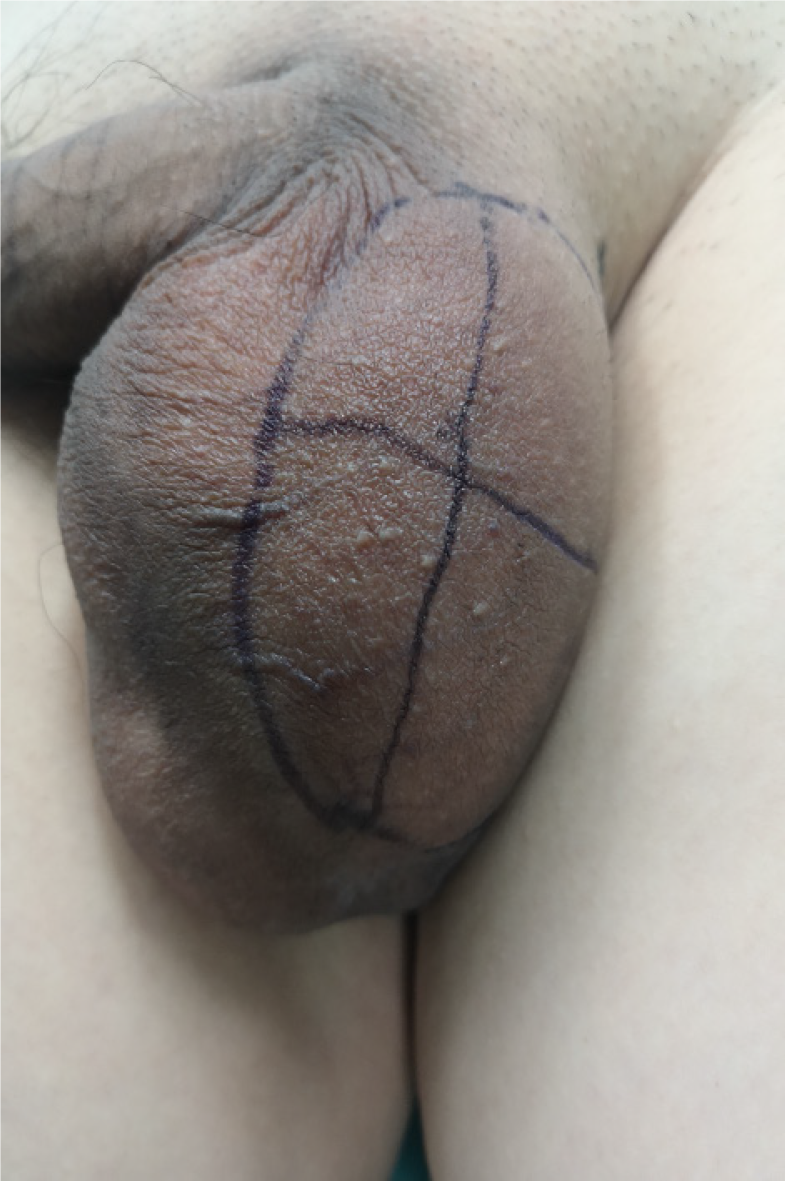
Physical examinations revealed a palpable subcutaneous mass measuring about 7.0 cm × 6.5 cm in the left scrotum, with tenderness (Figure 1). The left scrotum was enlarged, containing an oval-shaped mass with a smooth surface and no tenderness. Using a light transmission test, the testes on both sides were found to be palpable.
Laboratory findings were within normal limits. Serum human chorionic gonadotropin and alpha-fetoprotein were in normal level.
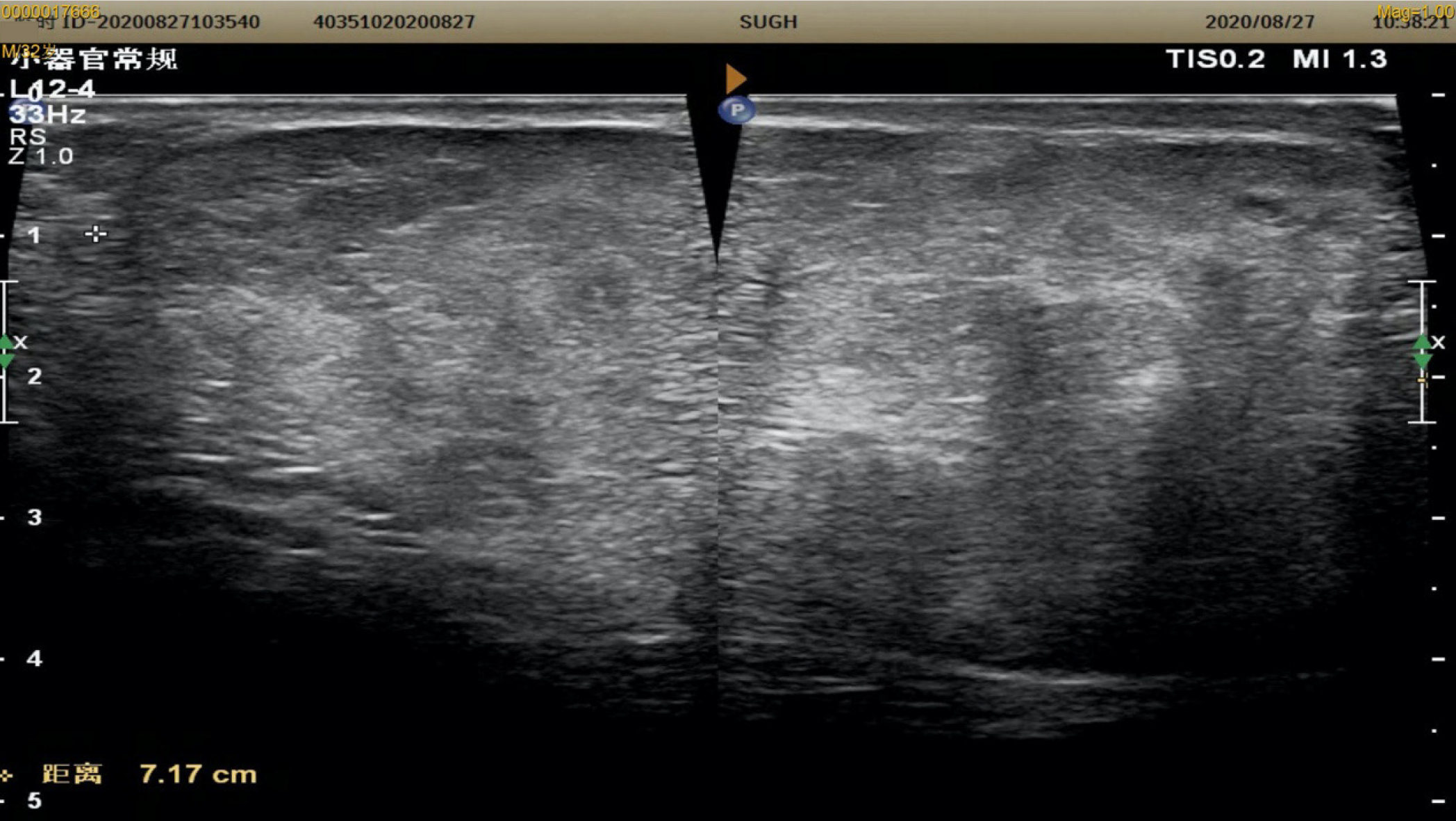
Color Doppler ultrasound of the scrotum, bilateral testes, and epididymis was performed, which revealed a subcutaneous hyperechoic mass in the left scrotum with a size of approximately 72 mm × 64 mm × 41 mm. The mass had a clear boundary, and the internal echo was uneven with visible sinusoids and strip-shaped blood flow signals. The mass was not connected to the abdominal cavity (Figure 2). No apparent abnormalities were observed in the other parts.
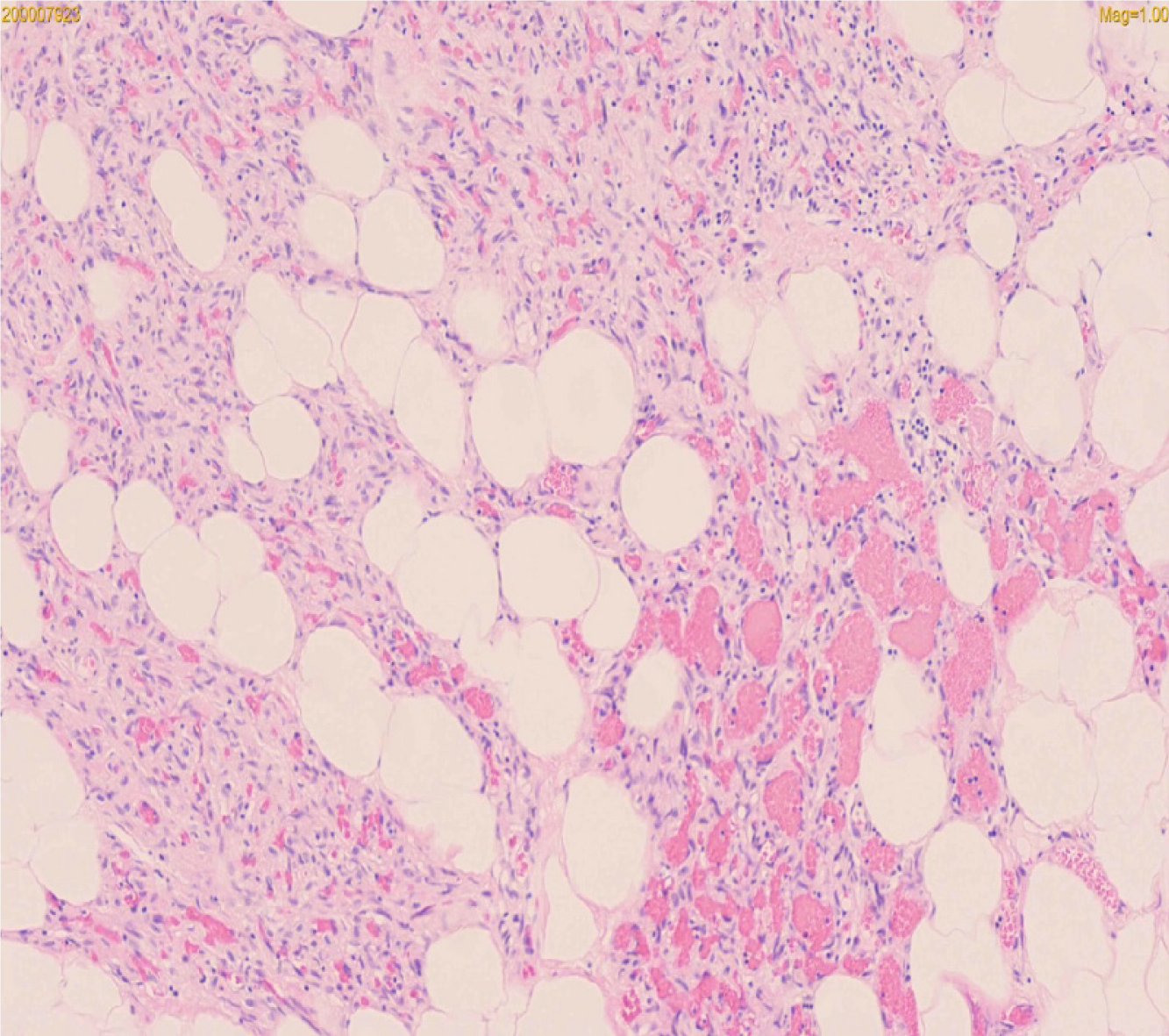
According to the results of pathology, the tumor was diagnosed as subcutaneous angiolipoma (Figure 3).
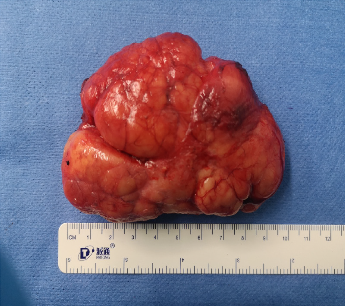
The patient underwent left scrotal mass resection (Figure 4).
After 4 d of treatment after the surgery, the patient was discharged. He was found to recover well at the 1-, 3-, and 6-mo follow-up after discharge.
Angiolipomas, first described by Bowen in 1912, account for 6%-17% of all lipomas[3]. They are benign mesenchymal tumors composed of lipoma tissue, capillaries, and stroma, with a vascular component of 15%-50%. This contrasts with the proportion of blood vessels in common lipomas, which is less than 10%[4]. Angiolipomas usually occur in teenagers and young adults between 20 and 25 years old, most commonly in the forearm (approximately two-thirds of cases), followed by the upper arm, thigh, and front abdominal wall, with a size of 1-4 cm. Men are more often affected than women[5]. Arenaz Bua found that 5% of the cases had a family heredity, and the specific inheritance pattern was uncertain. Tumours are often painful or tender and are most commonly seen in young men[6]. Another study found that adipose tissue tumours may be related to a history of trauma in addition to multiple familial lipomatosis and other genetic predispositions[7]. Clinical signs and symptoms are atypical and primarily manifest as multiple subcutaneous nodules, which may be accompanied by pain. Early lumps are small and often difficult to find because there are no obvious symptoms; when the lumps slowly increase in size, local symptoms of compression may occur.
We report the case of a 32-year-old man with an angiolipoma in the left scrotum. The left scrotum is an extremely rare site affected by angiolipomas. After reviewing the literature, we found that this is the first report of left scrotum angiolipoma with a length of more than 7 cm. Most para-testicular masses are benign, and lipomas account for approximately 90% of benign tumours in the para-testicular soft tissue[8]. The patient had no history of surgical resection, no history of trauma, and no family genetic history, and the pathological findings of this postoperative period were all angiolipomas. This case had unique characteristics in terms of location, size, management, and diagnosis.
Preoperative physical examinations cannot establish a diagnosis of angiolipomas, and ultrasound examination is the first choice of imaging modalities. Diagnosis must be confirmed by postoperative pathological examination. Therefore, the treatment of this disease should be based on surgery. A quick-frozen pathological examination during the operation should be performed to determine the nature of the disease. It should be distinguished from the lipomas or sarcomas, which have malignant or aggressive clinical courses.
Angiolipoma is a rare type of benign tumor extremely rarely found in the scrotum but needs to be considered when evaluating scrotal masses. Due to the economy and ease of use of ultrasound examinations, as well as the sensitivity to lesion location and size, it is the imaging test of choice for the characterisation of pathological masses in the scrotum and testes[9]. However, ultrasound is less specific in distinguishing between benign and malignant tumours[10]. Therefore, it is necessary to diagnose testicular angiolipoma through the histological evaluation of surgical specimens to determine the appropriate treatment plan.
We are grateful to the patient for providing relevant information and informed consent for publication of this case report.
Manuscript source: Unsolicited manuscript
Specialty type: Medicine, research and experimental
Country/Territory of origin: China
Peer-review report’s scientific quality classification
Grade A (Excellent): 0
Grade B (Very good): B
Grade C (Good): 0
Grade D (Fair): 0
Grade E (Poor): 0
P-Reviewer: Patel L S-Editor: Wang LL L-Editor: Wang TQ P-Editor: Li JH
| 1. | Lin JJ, Lin F. Two entities in angiolipoma. A study of 459 cases of lipoma with review of literature on infiltrating angiolipoma. Cancer. 1974;34:720-727. [RCA] [PubMed] [DOI] [Full Text] [Cited by in RCA: 1] [Reference Citation Analysis (0)] |
| 2. | Zhang Y, Yang J, Xu A, Li D, Yang G. A rare case of scrotal angiolipoma and the literature review. Urol Case Rep. 2020;32:101217. [RCA] [PubMed] [DOI] [Full Text] [Full Text (PDF)] [Cited by in Crossref: 1] [Cited by in RCA: 1] [Article Influence: 0.2] [Reference Citation Analysis (0)] |
| 3. | Altug HA, Sahin S, Sencimen M, Dogan N, Erdogan O. Non-infiltrating angiolipoma of the cheek: a case report and review of the literature. J Oral Sci. 2009;51:137-139. [RCA] [PubMed] [DOI] [Full Text] [Cited by in Crossref: 15] [Cited by in RCA: 19] [Article Influence: 1.2] [Reference Citation Analysis (0)] |
| 4. | Räsänen O, Nohteri H, Dammert K. Angiolipoma and lipoma. Acta Chir Scand. 1967;133:461-465. [PubMed] |
| 5. | Pattipati S, Kumar MN, Ramadevi, Kumar BP. Palatal lipoma: a case report. J Clin Diagn Res. 2013;7:3105-3106. [RCA] [PubMed] [DOI] [Full Text] [Cited by in Crossref: 2] [Cited by in RCA: 4] [Article Influence: 0.3] [Reference Citation Analysis (0)] |
| 6. | Garib G, Siegal GP, Andea AA. Autosomal-dominant familial angiolipomatosis. Cutis. 2015;95:E26-E29. [PubMed] |
| 7. | Copcu E, Sivrioglu NS. Posttraumatic lipoma: analysis of 10 cases and explanation of possible mechanisms. Dermatol Surg. 2003;29:215-220. [RCA] [PubMed] [DOI] [Full Text] [Cited by in Crossref: 19] [Cited by in RCA: 32] [Article Influence: 1.5] [Reference Citation Analysis (0)] |
| 8. | Başal Ş, Malkoç E, Aydur E, Yıldırım İ, Kibar Y, Kurt B, Göktaş S. Fibrous pseudotumors of the testis: The balance between sparing the testis and preoperative diagnostic difficulty. Turk J Urol. 2014;40:125-129. [RCA] [PubMed] [DOI] [Full Text] [Cited by in Crossref: 10] [Cited by in RCA: 10] [Article Influence: 1.0] [Reference Citation Analysis (0)] |
| 9. | Dogra VS, Gottlieb RH, Oka M, Rubens DJ. Sonography of the scrotum. Radiology. 2003;227:18-36. [RCA] [PubMed] [DOI] [Full Text] [Cited by in Crossref: 493] [Cited by in RCA: 397] [Article Influence: 18.0] [Reference Citation Analysis (0)] |
| 10. | Auer T, De Zordo T, Dejaco C, Gruber L, Pichler R, Jaschke W, Dogra VS, Aigner F. Value of Multiparametric US in the Assessment of Intratesticular Lesions. Radiology. 2017;285:640-649. [RCA] [PubMed] [DOI] [Full Text] [Cited by in Crossref: 40] [Cited by in RCA: 51] [Article Influence: 6.4] [Reference Citation Analysis (0)] |


