Copyright
©The Author(s) 2021.
World J Clin Cases. Jul 16, 2021; 9(20): 5709-5716
Published online Jul 16, 2021. doi: 10.12998/wjcc.v9.i20.5709
Published online Jul 16, 2021. doi: 10.12998/wjcc.v9.i20.5709
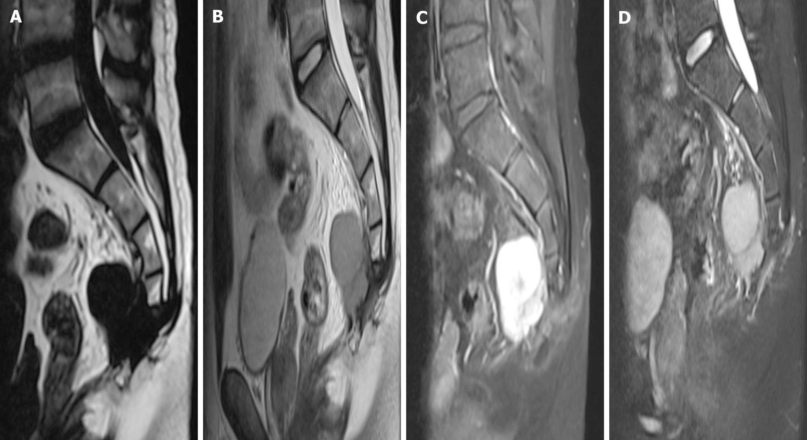
Figure 4 Magnetic resonance images showed an interval increase in the lesion with presacral extension, with low T1, high T2 signal, and avid enhancement.
A: Second preoperative T1-weighted sagittal magnetic resonance (MR) images; B: Second preoperative T2-weighted sagittal MR images; C: Second preoperative contrast-enhanced T1-weighted MR images; D: Second preoperative contrast-enhanced T2-weighted MR images.
- Citation: Zheng BW, Niu HQ, Wang XB, Li J. Sacral chondroblastoma — a rare location, a rare pathology: A case report and review of literature. World J Clin Cases 2021; 9(20): 5709-5716
- URL: https://www.wjgnet.com/2307-8960/full/v9/i20/5709.htm
- DOI: https://dx.doi.org/10.12998/wjcc.v9.i20.5709


