Published online Jul 6, 2021. doi: 10.12998/wjcc.v9.i19.5232
Peer-review started: January 18, 2021
First decision: February 11, 2021
Revised: February 18, 2021
Accepted: May 15, 2021
Article in press: May 15, 2021
Published online: July 6, 2021
Processing time: 156 Days and 19.4 Hours
Jejunal diverticula are the rarest of all small bowel diverticula and usually have no classic clinical symptoms. Jejunal diverticular haemorrhage (JDH) is a rare complication and can be difficult to identify and manage, hence it always resulting in a diagnostic delay and unsatisfactory clinical outcomes. Although with the advances in endoscopic technology, no consensus have been reached on the diagnosis and management of JDH, the conventional surgical intervention still remains the mainstream for the management of JDH. We report an unique case of a 63-year-old male who presented with massive haemorrhage from jejunal diverticula, which was successfully managed by initial resuscitation and defi
A 63-year-old male was admitted as an emergency with 6 h history of haema
In patients with gastrointestinal bleeding, if various techniques fail to identify the cause of haemorrhage in small bowel and haemodynamic instability is sustained with continuous resuscitation, we recommend surgical intervention should be the ultimate treatment of choice.
Core Tip: In patients with gastrointestinal bleeding, if all methods have failed to identify the cause of haemorrhage in small bowel and haemodynamic instability sustains with continuous resuscitation, we recommend surgical intervention as the ultimate treatment of choice for jejunal diverticular haemorrhage. Surgeons should strictly follow the diagnosis and treatment guidelines of acute gastrointestinal bleeding and have a better understanding of the strengths and weaknesses of various techniques, which would be extremely helpful for selecting the optimal clinical pathways and conducting multidisciplinary collaboration accurately and quickly.
- Citation: Ma HC, Xiao H, Qu H, Wang ZJ. Successful diagnosis and treatment of jejunal diverticular haemorrhage by full-thickness enterotomy: A case report. World J Clin Cases 2021; 9(19): 5232-5237
- URL: https://www.wjgnet.com/2307-8960/full/v9/i19/5232.htm
- DOI: https://dx.doi.org/10.12998/wjcc.v9.i19.5232
Small intestinal diverticula are quite rare, with incidence ranges from 0.06% to 1.3%, and a slight male predominance[1]. All diverticula are usually acquired except for Meckel's diverticulum. The most common site of small intestinal diverticulosis is the duodenum (60%-70%), then followed by jejunum (20%-25%) and ileum (5%-10%). Jejunal diverticula are commonly found in the proximal jejunum and manifested with multiple localizations. The formation of diverticula is the result of herniation of the mucosa, submucosa, and serosa through the muscular layer of the bowel at the point where the vasa recta enter the muscularis propria[2].
The presentation of jejunal diverticulosis is variable, from asymptomatic to chronic abdominal symptoms, and about 10%-30% of patients with jejunoileal diverticula will develop acute complications such as acute abdominal pain, haemorrhage, diver
A 63-year-old male was admitted to the emergency department with 6 h history of haematemesis and melena.
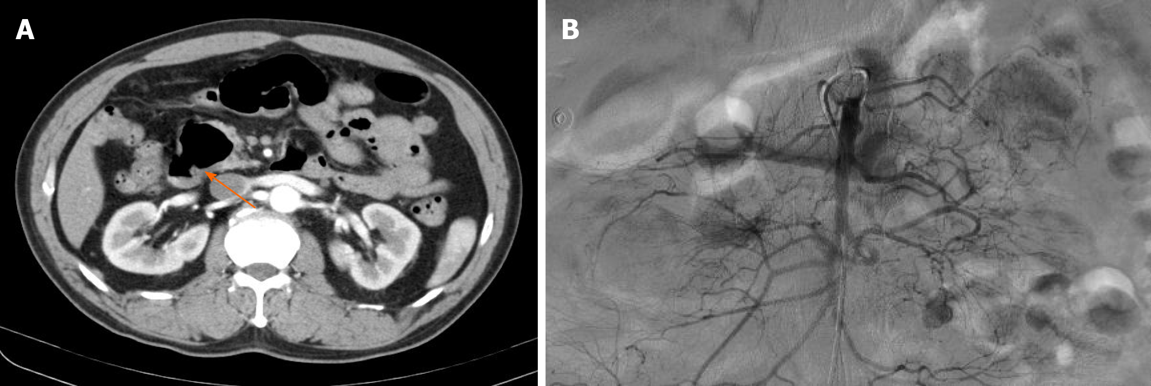
The patient had a symptom of haematemesis and melena for 6 h. The haematemesis appeared to be bright red, with volume exceeding 100 mL. The amount of melena was estimated to be 200 mL. He was admitted into our emergency department. He was haemodynamically unstable with a soft, non-tender abdomen. His haemoglobin was 5.2 g/dL, and white blood cell count was 12.0 cells/mm3. Computed tomography (CT) scanning revealed that there were dilated small bowel loops with multiple jejunal diverticula. For further treatment, the patient was transferred to our general surgery department.
No special past medical treatment history such as corticosteroids, thrombolytic therapy, and anticoagulation.
He was retired and a current smoker (10 cigarettes/d for the past 30 years). He had no serious family history.
Physical examination showed abdominal tenderness in the whole abdomen, and muscle tension was not palpated. The bowel sounded active, with six to eight bowel sounds per min. Rectal examination revealed dark blood with no masses.
His hemoglobin was 5.2 g/dL, and white blood cell count was 12.0 cells/mm3.
The abdominal CT showed dilated small bowel loops with multiple jejunal diverticula (Figure 1A).
JDH, acute haemorrhagic shock.
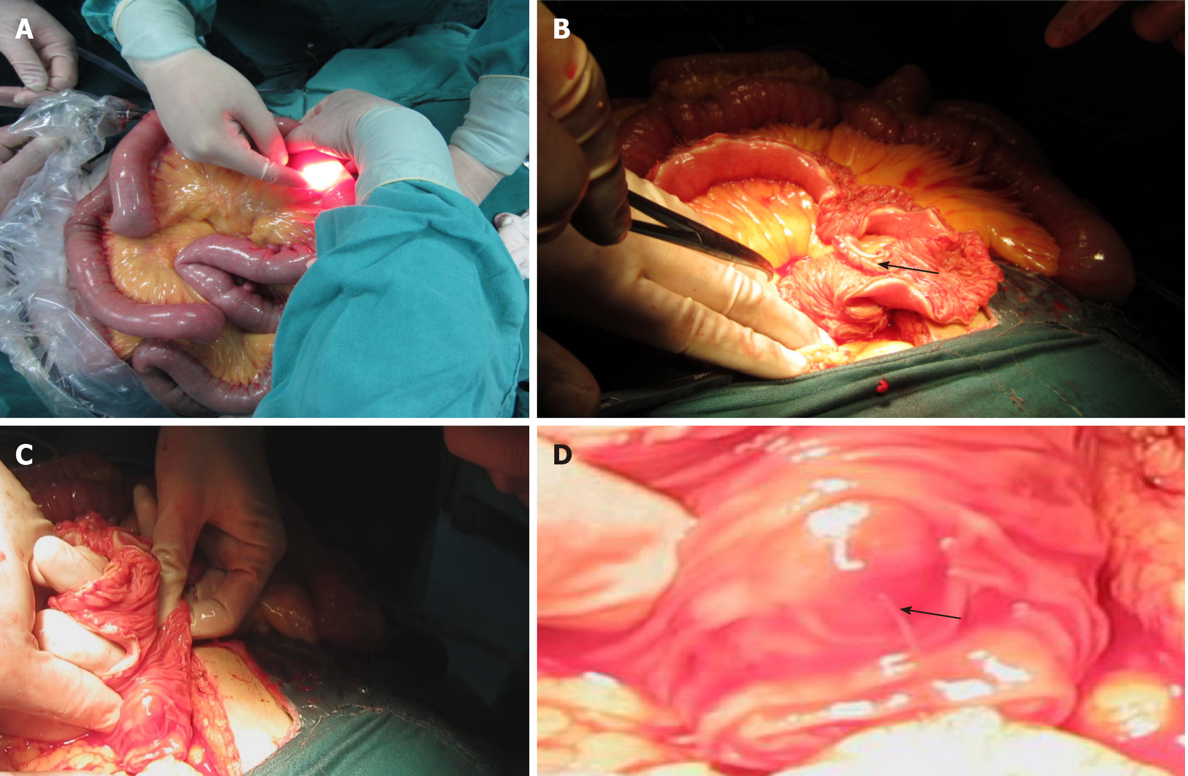
After fluid resuscitation and three unit blood transfusion, he had a normal upper gastrointestinal endoscopy but without positive findings. Colonoscopy only showed dark blood but no obvious bleeding source. The mesenteric angiography was performed subsequently; also no visible sites of bleeding were shown (Figure 1B). The symptoms of haematemesis and melena became aggravated during the period of examination, which indicated progressive bleeding. Without hesitation, an exploratory laparotomy was performed. Written informed consent was obtained from the patient. A rapid exploration revealed multiple jejunal diverticulum 60 cm from the duodeno-jejunal flexure. However, we found it impossible to detect the small bowel lesions by palpation. The gastroscope was inserted into the lumen via a small incision in jejunum (Figure 2A), which only showed extensive red blood but did not identify the pulsating bleeding site. The endoscope failed to provide an excellent visualization due to the large amount of blood in the jejunum. We had no alternative but to make a 60 cm length jejunum incision longitudinally in the antimesenteric border (Figure 2B), the diverticula were detected from proximal to distal under direct vision. Finally, a pulsating vessel was identified in the first diverticulum under the duodeno-jejunal flexure (Figure 2C and Figure 2D). Haemorrhage stopped through suture and ligation of the bleeding site. A segmental jejunal resection and a functional side-to-side stapled anastomosis were conducted.
The postoperative period was uneventful, and he was discharged on day 18 after surgery. At the 1-year follow-up, no rebleeding has occurred.
Small intestinal diverticula are very rare and were firstly described in 1794 by Somerling. Jejunal diverticula are the rarest of all small bowel diverticula[4]. The majority of patients with small bowel diverticula are usually asymptomatic, but 10%-30% patients may encounter chronic abdominal aspecific symptoms and acute complications such as bleeding, perforation, and obstruction[5]. A recent study involving 527 patients with jejunoileal diverticula reported that haemorrhage was the most frequent complication (30%), followed by perforation (23%), obstruction (17.3%), and non-complicated acute diverticulitis (14.9%)[6].
Braithwaite[7] reported the first case of bleeding from jejunal diverticulosis in 1923. JDH is an unusual cause of severe small bowel bleeding. However, it could be fatal for patients. Haemorrhage from jejunal diverticula mainly manifest with lower gastrointestinal bleeding, although cases of haematemesis have been reported occasionally[8]. The haemorrhage within jejunal diverticula can be attributed to the following reasons: (1) Bleeding occurred when vessels were corroded by diverticulitis with ulceration; (2) Haemorrhage caused by erosive effects of digestive fluids when there existed ectopic gastric mucosa or pancreatic tissue in the diverticulum; and (3) Bleeding aroused by the administration of anticoagulants such as aspirin and warfarin.
How to identify the bleeding site in the small bowel remains a clinically challenging issue. Small bowel haemorrhage has multiple factors, including tumors, diverticula, ulcers, vessel malformations, inflammatory diseases, etc. Conventionally, it can be visualized with CT and treated surgically for the large lesion in the small bowel. However, the causes and sites of lesions of haemorrhage from a minor or a flat lesion in the small intestine are sometimes extremely difficult to identify using conventional approaches such as endoscopy, radiography, angiography and scintigraphy.
Massive bleeding in small bowel that lead to shock is not rarely seen clinically, which usually required emergency surgery. However, it may be troublesome for localization of the lesion intraoperative if it is too small or soft to be palpated from the serosal side. Although various techniques have been attempted, including preo
No consensus has been reached in the diagnosis and treatment of complicated jejunal diverticula so far. Various strategies have been adopted and largely depend on personal experience and skills. According to our experiences in the management of JDH, the abdominal CT is initially utilized to detect the cause of bleeding. Although abdominal CT may show small bowel diverticula clearly, it is not sensitive enough to detect haemorrhagic lesions. For the patients with gastrointestinal bleeding, the diagnostic endoscopy, including upper gastrointestinal endoscopy and colonoscopy, should be performed. Colonic and gastroesophageal lesions can be detected easily through colonoscopy and gastrointestinal endoscopy.
Radionuclide imaging and angiography may be appropriate in patients with massive gastrointestinal haemorrhage in whom the bleeding source has not been identified through colonoscopy and gastrointestinal endoscopy. However, the angiography might identify a haemorrhage site only when the rate of ongoing arterial bleeding is more than 1 mL/min[9]. Angiographic localization also allows for embolization for the patients with acute arterial bleeding. Angiography requires a bleeding rate > 1 mL/min for accurate detection of extravasation of contrast into the bowel lumen through radionuclide imaging using technetium tagged red blood cells. Delayed scans are unreliable for identification of the bleeding site in small bowel.
With the constant improvements in endoscopy technology, videocapsule endoscopy and double-balloon endoscopy were applied in the diagnosis of small intestinal lesions. As a noninvasive tool to explore small bowel, the diagnostic yield of video
In patients with gastrointestinal bleeding, if all of the above methods fail in identifying the cause of haemorrhage in small bowel and haemodynamic instability sustains although continuous resuscitation, we recommend surgical intervention as the ultimate treatment of choice for JDH. As illustrated by this case, we should infer the most likely bleeding site during the exploratory laparotomy, and then shrink the exploratory area gradually, localizing the bleeding site and preventing eventually the haemorrhage. However, if the bleeding source cannot be detected through intraoperative enteroscopy, the full-thickness enterotomy with exploration should be executed immediately.
Surgeons should strictly follow the diagnosis and treatment guidelines of acute gastrointestinal bleeding and have a better understanding of the strengths and weaknesses of various techniques. This would be extremely helpful for selecting the optimal clinical pathways and conducting multidisciplinary collaboration accurately and quickly.
The manuscript was prepared and revised according to the CARE Checklist (2016).
Manuscript source: Unsolicited manuscript
Specialty type: Surgery
Country/Territory of origin: China
Peer-review report’s scientific quality classification
Grade A (Excellent): 0
Grade B (Very good): 0
Grade C (Good): C
Grade D (Fair): 0
Grade E (Poor): 0
P-Reviewer: Thosani N S-Editor: Ma YJ L-Editor: Filipodia P-Editor: Wang LL
| 1. | Akhrass R, Yaffe MB, Fischer C, Ponsky J, Shuck JM. Small-bowel diverticulosis: perceptions and reality. J Am Coll Surg. 1997;184:383-388. [RCA] [PubMed] [DOI] [Full Text] [Cited by in Crossref: 41] [Cited by in RCA: 38] [Article Influence: 1.4] [Reference Citation Analysis (0)] |
| 2. | Fidan N, Mermi EU, Acay MB, Murat M, Zobaci E. Jejunal Diverticulosis Presented with Acute Abdomen and Diverticulitis Complication: A Case Report. Pol J Radiol. 2015;80:532-535. [RCA] [PubMed] [DOI] [Full Text] [Full Text (PDF)] [Cited by in Crossref: 7] [Cited by in RCA: 9] [Article Influence: 0.9] [Reference Citation Analysis (0)] |
| 3. | Butler JS, Collins CG, McEntee GP. Perforated jejunal diverticula: a case report. J Med Case Rep. 2010;4:172. [RCA] [PubMed] [DOI] [Full Text] [Full Text (PDF)] [Cited by in Crossref: 27] [Cited by in RCA: 35] [Article Influence: 2.3] [Reference Citation Analysis (0)] |
| 4. | Bellio G, Kurihara H, Zago M, Tartaglia D, Chiarugi M, Coppola S, Biloslavo A, de Manzini N. Jejunoileal diverticula: a broad spectrum of complications. ANZ J Surg. 2020;90:1454-1458. [RCA] [PubMed] [DOI] [Full Text] [Cited by in Crossref: 3] [Cited by in RCA: 4] [Article Influence: 0.8] [Reference Citation Analysis (0)] |
| 5. | Nonose R, Valenciano JS, de Souza Lima JS, Nascimento EF, Silva CM, Martinez CA. Jejunal Diverticular Perforation due to Enterolith. Case Rep Gastroenterol. 2011;5:445-451. [RCA] [PubMed] [DOI] [Full Text] [Full Text (PDF)] [Cited by in Crossref: 19] [Cited by in RCA: 18] [Article Influence: 1.3] [Reference Citation Analysis (0)] |
| 6. | Abongwa HK, De Simone B, Alberici L, Iaria M, Perrone G, Tarasconi A, Baiocchi G, Portolani N, Di Saverio S, Sartelli M, Coccolini F, Manegold JE, Ansaloni L, Catena F. Implications of Left-sided Gallbladder in the Emergency Setting: Retrospective Review and Top Tips for Safe Laparoscopic Cholecystectomy. Surg Laparosc Endosc Percutan Tech. 2017;27:220-227. [RCA] [PubMed] [DOI] [Full Text] [Cited by in Crossref: 19] [Cited by in RCA: 16] [Article Influence: 2.0] [Reference Citation Analysis (0)] |
| 7. | Braithwaite LR. A case of jejunal diverticula. Br J Surg. 1923;11:184-8. [RCA] [DOI] [Full Text] [Cited by in Crossref: 16] [Cited by in RCA: 15] [Article Influence: 0.8] [Reference Citation Analysis (0)] |
| 8. | Altemeier WA, Bryant LR, Wulsin JH. The surgical significance of jejunal diverticulosis. Arch Surg. 1963;86:732-745. [RCA] [PubMed] [DOI] [Full Text] [Cited by in Crossref: 73] [Cited by in RCA: 69] [Article Influence: 2.6] [Reference Citation Analysis (0)] |
| 9. | Edelman DA, Sugawa C. Lower gastrointestinal bleeding: a review. Surg Endosc. 2007;21:514-520. [RCA] [PubMed] [DOI] [Full Text] [Cited by in Crossref: 55] [Cited by in RCA: 29] [Article Influence: 1.6] [Reference Citation Analysis (0)] |
| 10. | Koffas A, Laskaratos FM, Epstein O. Non-small bowel lesion detection at small bowel capsule endoscopy: A comprehensive literature review. World J Clin Cases. 2018;6:901-907. [RCA] [PubMed] [DOI] [Full Text] [Full Text (PDF)] [Cited by in CrossRef: 9] [Cited by in RCA: 9] [Article Influence: 1.3] [Reference Citation Analysis (0)] |


