Published online Jun 26, 2021. doi: 10.12998/wjcc.v9.i18.4760
Peer-review started: December 16, 2020
First decision: January 10, 2021
Revised: January 14, 2021
Accepted: April 23, 2021
Article in press: April 23, 2021
Published online: June 26, 2021
Processing time: 176 Days and 18.9 Hours
Clinical femoral neck fracture is common. Based on patient age and fracture type, different surgical methods can be selected, including cannulated screw fixation of the femoral neck and artificial total hip joint or semi-hip joint replacement. When patients with femoral neck fracture are treated with cannulated screw fixation, a cannulated screw may be positioned too deep. The excessively deep-placed screw is difficult to remove and causes major trauma to the patient.
A patient with poliomyelitis and femoral neck fracture was treated with a cannulated screw that was placed too deep. A self-made auxiliary tool (made of a steel sternal wire) was used to remove the cannulated screw near the pelvic cavity.
The depth of the cannulated screw can be estimated before screw placement using an improved hollow screwdriver with a scale mark, thus improving the safety of screw placement and facilitating clinical use.
Core Tip: A patient with poliomyelitis and femoral neck fracture was treated with a cannulated screw that was placed too deep. A self-made auxiliary tool (made of a steel sternal wire) was used to remove the cannulated screw near the pelvic cavity. The depth of the cannulated screw can be estimated before screw placement using an improved hollow screwdriver with a scale mark.
- Citation: Yang ZH, Hou FS, Yin YS, Zhao L, Liang X. Minimally invasive removal of a deep-positioned cannulated screw from the femoral neck: A case report. World J Clin Cases 2021; 9(18): 4760-4764
- URL: https://www.wjgnet.com/2307-8960/full/v9/i18/4760.htm
- DOI: https://dx.doi.org/10.12998/wjcc.v9.i18.4760
Clinical femoral neck fracture is common[1]. Patients with femoral neck fracture can be treated with cannulated screw fixation, but a cannulated screw may be positioned too deep in clinical practice. The excessively deep-placed screw is difficult to remove and causes major trauma to the patient.
A 59-year-old Chinese man was hospitalized in the Department of Orthopaedics at the Second Hospital of Shanxi Medical University (Taiyuan, China) because of pain in his left hip.
In 1995, the patient underwent orthopedic surgery for poliomyelitis performed by an unknown surgeon.
The patient did not smoke or drink. He had no history of other illnesses and had no known allergies.
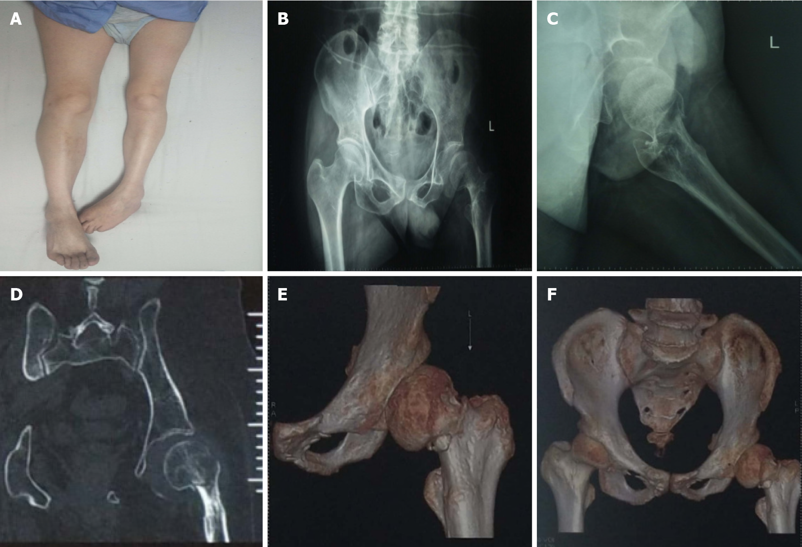
His medical history included poliomyelitis[2] with limited activity of the left hip joint. The left limb was shorter than the right.
Films taken in the local hospital revealed a Garden II[3] left femoral neck fracture, and preoperative computed tomography indicated a left femoral neck fracture (Figure 1).
Based on the examination and imaging findings, a provisional diagnosis of left femoral neck fracture and poliomyelitis was made.
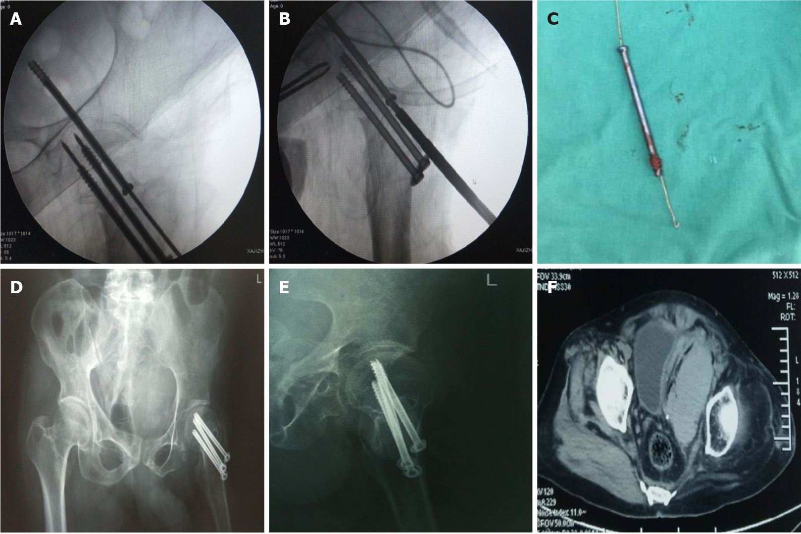
With the patient’s age and basic diseases known, closed reduction of cannulated screw internal fixation for femoral neck fracture was performed[4]. Intraoperative fluoroscopic examination using the traction bed revealed good reduction. A cannulated screw guide pin was positioned percutaneously via minimally invasive surgery[5], and intraoperative fluoroscopy showed that the length of the guide pin was appropriate. A screw with appropriate length with respect to the measured depth was inserted, but intraoperative fluoroscopy showed that the upper cannulated screw was placed too deep and penetrated the acetabulum[6]. After attempts to unscrew the screw failed, the closed incision was connected and expanded to reach the bone. The screw tail completely entered the bone, and the cannulated screw could not be taken out by retrograde rotation of the screwdriver. An auxiliary extraction tool was made by bending the steel sternal wire and then pushing it through the rear of the screwdriver to reach the tip of the cannulated screw. The screwdriver was rotated reversely[7], and the steel sternal wire was pulled gently. The overlong hollow screw was slowly rotated out and replaced with a thickened hollow screw of appropriate length and a gasket implanted into the femoral neck. After surgery, the wound was washed thoroughly and sutured layer by layer. The patient was advised to stop eating temporarily. Bedside B-mode ultrasound revealed a heterogeneous mass on the left side of the bladder, with no obvious abnormality. A pelvic computed tomography scan showed effusion (Figure 2). A regular B-mode ultrasound examination showed normal urine and stool[8], and the patient was discharged 1 wk after surgery.
Re-examination 2 years after surgery showed that the patient’s urine and stools were normal, and the fracture was healed. Abdominal ultrasound showed no obvious abnormality, and the activity of the hip joint recovered to preoperative levels.
Based on patient age and fracture type, different surgical methods can be selected for femoral neck fracture, including cannulated screw fixation of the femoral neck and artificial total hip joint or semi-hip joint replacement[9]. This patient had underlying poliomyelitis, and his left hip joint had limited daily activity. Poliomyelitis leads to poor hip muscle strength, and thus the strategy of closed reduction and cannulated screw placement was adopted. However, the screw placement was difficult due to osteoporosis. Less intraoperative fluoroscopy was performed, leading to excessively deep insertion of the screw. The cannulated screw penetrated the femoral head and the ipsilateral acetabulum, and it was difficult to unscrew directly by reverse rotation because of no bone or thread friction.
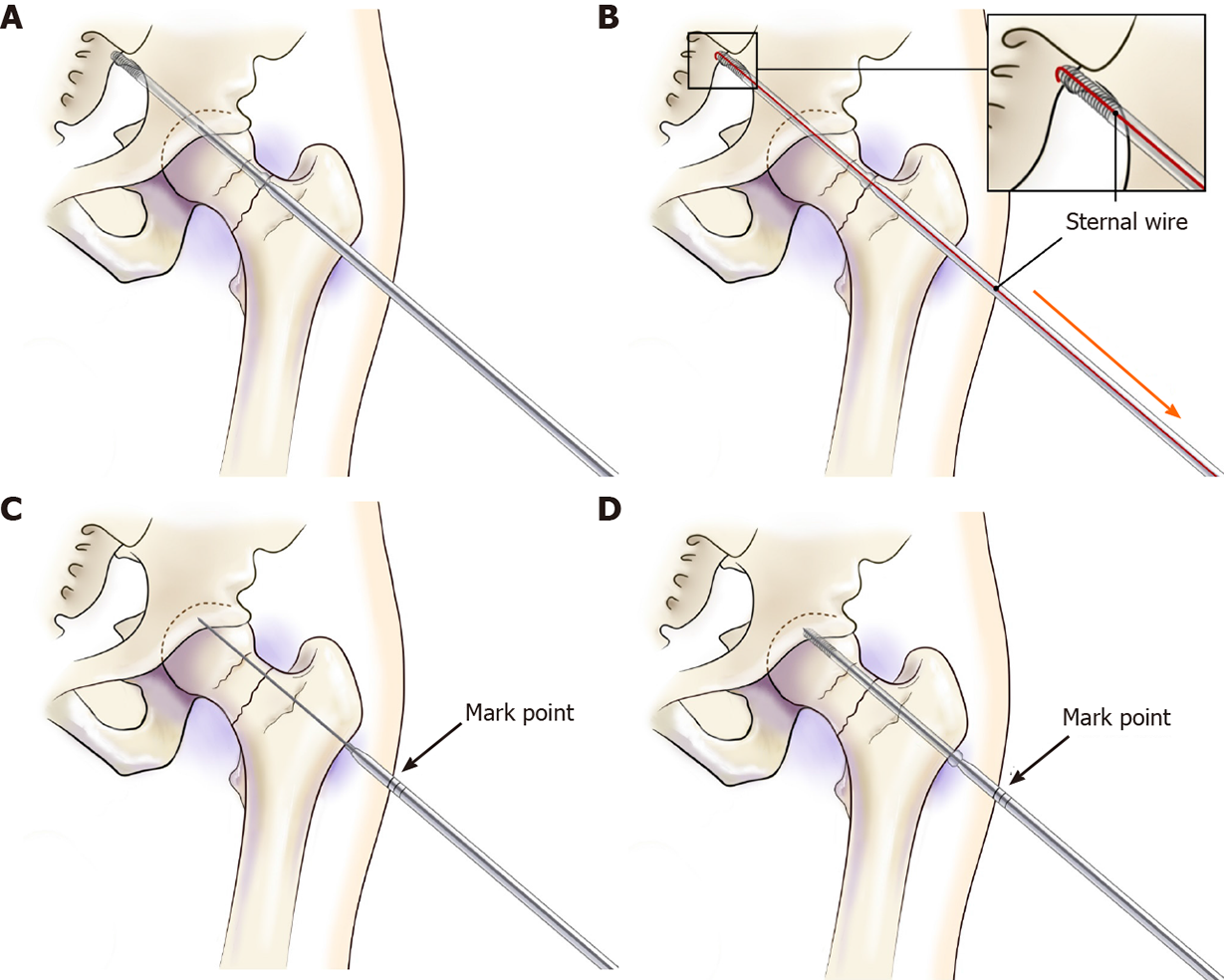
The Stoppa approach[10] and ilioinguinal approach can be used for treatment of acetabular fractures. Surgery can reach the top of the acetabulum and remove the cannulated screw. However, there is major surgical trauma for patients. The tool made of the thickest steel sternal wire, with a small bend at its head, facilitated rotation of the screwdriver to remove the cannulated screw, which avoided cutting via the ilioinguinal approach[11] or Stoppa approach to remove the overly deep-positioned screw. Regarding its usage, a guide pin was put on the screwdriver to reach the fracture before inserting the cannulated screw, and the scale of the screwdriver and the mark at its interface with the skin were observed. After the cannulated screw was inserted, the depth was determined by fluoroscopy when the mark on the skin and the scale of the original measuring screwdriver were aligned (Figure 3), reducing intraoperative fluoroscopy and surgical time and enhancing the safety of screw positioning.
The depth of the cannulated screw can be estimated before screw placement using an improved hollow screwdriver with a scale mark.
Manuscript source: Unsolicited manuscript
Specialty type: Medicine, research and experimental
Country/Territory of origin: China
Peer-review report’s scientific quality classification
Grade A (Excellent): 0
Grade B (Very good): B
Grade C (Good): 0
Grade D (Fair): 0
Grade E (Poor): 0
P-Reviewer: Toro G S-Editor: Zhang L L-Editor: Filipodia P-Editor: Wu YXJ
| 1. | Lu Y, Wang Y, Song Z, Wang Q, Sun L, Ren C, Xue H, Li Z, Zhang K, Hao D, Zhao Y, Ma T. Treatment comparison of femoral shaft with femoral neck fracture: a meta-analysis. J Orthop Surg Res. 2020;15:19. [RCA] [PubMed] [DOI] [Full Text] [Full Text (PDF)] [Cited by in Crossref: 3] [Cited by in RCA: 3] [Article Influence: 0.6] [Reference Citation Analysis (0)] |
| 2. | Chang KH, Lai CH, Chen SC, Tang IN, Hsiao WT, Liou TH, Lee CM. Femoral neck bone mineral density in ambulatory men with poliomyelitis. Osteoporos Int. 2011;22:195-200. [RCA] [PubMed] [DOI] [Full Text] [Cited by in Crossref: 26] [Cited by in RCA: 19] [Article Influence: 1.4] [Reference Citation Analysis (0)] |
| 3. | Lapidus LJ, Charalampidis A, Rundgren J, Enocson A. Internal fixation of garden I and II femoral neck fractures: posterior tilt did not influence the reoperation rate in 382 consecutive hips followed for a minimum of 5 years. J Orthop Trauma. 2013;27:386-90; discussion 390. [RCA] [PubMed] [DOI] [Full Text] [Cited by in Crossref: 41] [Cited by in RCA: 47] [Article Influence: 3.9] [Reference Citation Analysis (0)] |
| 4. | Kakar S, Tornetta P 3rd, Schemitsch EH, Swiontkowski MF, Koval K, Hanson BP, Jönsson A, Bhandari M; International Hip Fracture Research Collaborative. Technical considerations in the operative management of femoral neck fractures in elderly patients: a multinational survey. J Trauma. 2007;63:641-646. [RCA] [PubMed] [DOI] [Full Text] [Cited by in Crossref: 26] [Cited by in RCA: 27] [Article Influence: 1.6] [Reference Citation Analysis (0)] |
| 5. | Kang HG, Roh YW, Kim HS. The treatment of metastasis to the femoral neck using percutaneous hollow perforated screws with cement augmentation. J Bone Joint Surg Br. 2009;91:1078-1082. [RCA] [PubMed] [DOI] [Full Text] [Cited by in Crossref: 15] [Cited by in RCA: 18] [Article Influence: 1.1] [Reference Citation Analysis (0)] |
| 6. | Silver JK, Aiello DD. Polio survivors: falls and subsequent injuries. Am J Phys Med Rehabil. 2002;81:567-570. [RCA] [PubMed] [DOI] [Full Text] [Cited by in Crossref: 52] [Cited by in RCA: 43] [Article Influence: 1.9] [Reference Citation Analysis (0)] |
| 7. | Toro G, Moretti A, Paoletta M, De Cicco A, Braile A, Panni AS. Neglected femoral neck fractures in cerebral palsy: a narrative review. EFORT Open Rev. 2020;5:58-64. [RCA] [PubMed] [DOI] [Full Text] [Full Text (PDF)] [Cited by in Crossref: 6] [Cited by in RCA: 5] [Article Influence: 1.0] [Reference Citation Analysis (0)] |
| 8. | Crouse JR 3rd. B-mode ultrasound in clinical trials. Answers and questions. Circulation. 1993;88:319-321. [RCA] [PubMed] [DOI] [Full Text] [Cited by in Crossref: 26] [Cited by in RCA: 29] [Article Influence: 0.9] [Reference Citation Analysis (0)] |
| 9. | Jagatia M, Jin ZM. Analysis of elastohydrodynamic lubrication in a novel metal-on-metal hip joint replacement. Proc Inst Mech Eng H. 2002;216:185-193. [RCA] [PubMed] [DOI] [Full Text] [Cited by in Crossref: 21] [Cited by in RCA: 21] [Article Influence: 0.9] [Reference Citation Analysis (0)] |
| 10. | Champault GG, Rizk N, Catheline JM, Turner R, Boutelier P. Inguinal hernia repair: totally preperitoneal laparoscopic approach vs Stoppa operation: randomized trial of 100 cases. Surg Laparosc Endosc. 1997;7:445-450. [RCA] [PubMed] [DOI] [Full Text] [Cited by in Crossref: 82] [Cited by in RCA: 64] [Article Influence: 2.4] [Reference Citation Analysis (0)] |
| 11. | Matta JM. Operative treatment of acetabular fractures through the ilioinguinal approach: a 10-year perspective. J Orthop Trauma. 2006;20:S20-S29. [RCA] [PubMed] [DOI] [Full Text] [Cited by in Crossref: 131] [Cited by in RCA: 118] [Article Influence: 6.2] [Reference Citation Analysis (0)] |


