Published online Jun 6, 2021. doi: 10.12998/wjcc.v9.i16.4095
Peer-review started: January 23, 2021
First decision: February 11, 2021
Revised: February 19, 2021
Accepted: March 15, 2021
Article in press: March 15, 2021
Published online: June 6, 2021
Processing time: 111 Days and 1.6 Hours
Arrhythmogenic right ventricular (RV) cardiomyopathy is a rare and currently underrecognized cardiomyopathy characterized by the replacement of RV myocardium by fibrofatty tissue. It may be asymptomatic or symptomatic (palpitations or syncope) and may induce sudden cardiac death, especially during exercise. To prevent adverse events such as sudden cardiac death and heart failure, early diagnosis and treatment of arrhythmogenic RV cardiomyopathy (ARVC) are crucial. We report a patient with ARVC characterized by recurrent syncope during exercise who was successfully treated with combined endocardial and epicardial catheter ablation.
A 43-year-old man was referred for an episode of syncope during exercise. Previously, the patient experienced two episodes of syncope without a firm etiological diagnosis. An electrocardiogram obtained at admission indicated ventricular tachycardia originating from the inferior wall of the right ventricle. The ventricular tachycardia was terminated with intravenous propafenone. A repeat electrocardiogram showed a regular sinus rhythm with negative T waves and a delayed S-wave upstroke from leads V1 to V4. Cardiac magnetic resonance imaging showed RV free wall thinning, regional RV akinesia, RV dilatation and fibrofatty infiltration (RV ejection fraction of 38%). An electrophysiological study showed multiple inducible ventricular tachycardia as of a focal mechanism from the right ventricle. Endocardial and epicardial voltage mapping demonstrated scar tissue in the anterior wall, free wall and posterior wall of the right ventricle. Late potentials were also recorded. The patient was diagnosed with ARVC and treated with combined endocardial and epicardial catheter ablation with a very satisfactory follow-up result.
Clinicians should be aware of ARVC, and further workup, including imaging with multiple modalities, should be pursued. The combination of epicardial and endocardial catheter ablation can lead to a good outcome.
Core Tip: Classification of cardiomyopathy based on clinical phenotype is critical. In general, arrhythmogenic right ventricular cardiomyopathy should be suspected in patients with right ventricular arrhythmia, a family history of arrhythmogenic right ventricular cardiomyopathy or sudden death or abnormal electrocardiogram features such as repolarization or depolarization abnormalities in the right leads. We hope that this case will raise awareness of arrhythmogenic right ventricular cardiomyopathy.
- Citation: Wu HY, Cao YW, Gao TJ, Fu JL, Liang L. Arrhythmogenic right ventricular cardiomyopathy characterized by recurrent syncope during exercise: A case report. World J Clin Cases 2021; 9(16): 4095-4103
- URL: https://www.wjgnet.com/2307-8960/full/v9/i16/4095.htm
- DOI: https://dx.doi.org/10.12998/wjcc.v9.i16.4095
Arrhythmogenic right ventricular cardiomyopathy (ARVC) is a rare and under
A 43-year-old man presented with recurrent episodes of syncope during exercise for 6 mo.
Previously, the patient had reported two episodes of syncope during exercise (running), 6 mo and 2 mo before the current presentation. The patient was referred to a local hospital, where a 12-lead ECG indicated ventricular tachycardia (VT). An intravenous amiodarone infusion was given, and the VT was terminated. The patient then took amiodarone regularly without further examination or treatment. One hour before admission to our hospital, the patient experienced an episode of syncope with a recovery time of approximately 2 min during exercise (running). The patient recovered spontaneously without cardiopulmonary resuscitation. Subsequently, the patient felt persistent palpitations.
The patient had a 4-year history of hypertension and was treated by nifedipine controlled-release tablets (30 mg/d) with blood pressure controlled at approximately 130/80 mmHg.
The patient had no relevant personal history. The patient denied a family history of premature coronary artery disease or sudden cardiac death. Genetically inherited cardiomyopathies or arrhythmias were also denied.
The patient’s vital signs on arrival were as follows: Blood pressure of 110/68 mmHg, heart rate 192 beats per minute and respiratory rate of 22 breaths per minute. Pulmonary and cardiac examinations showed no significant abnormalities. Jugular vein engorgement and peripheral edema were not found.
The troponin I level was 0.78 ng/mL (normal range < 0.04). The patient’s leukocyte, hemoglobin, inflammatory factor, renal function, liver function, electrolyte, D-dimer and B-type natriuretic peptide levels were not significantly abnormal.
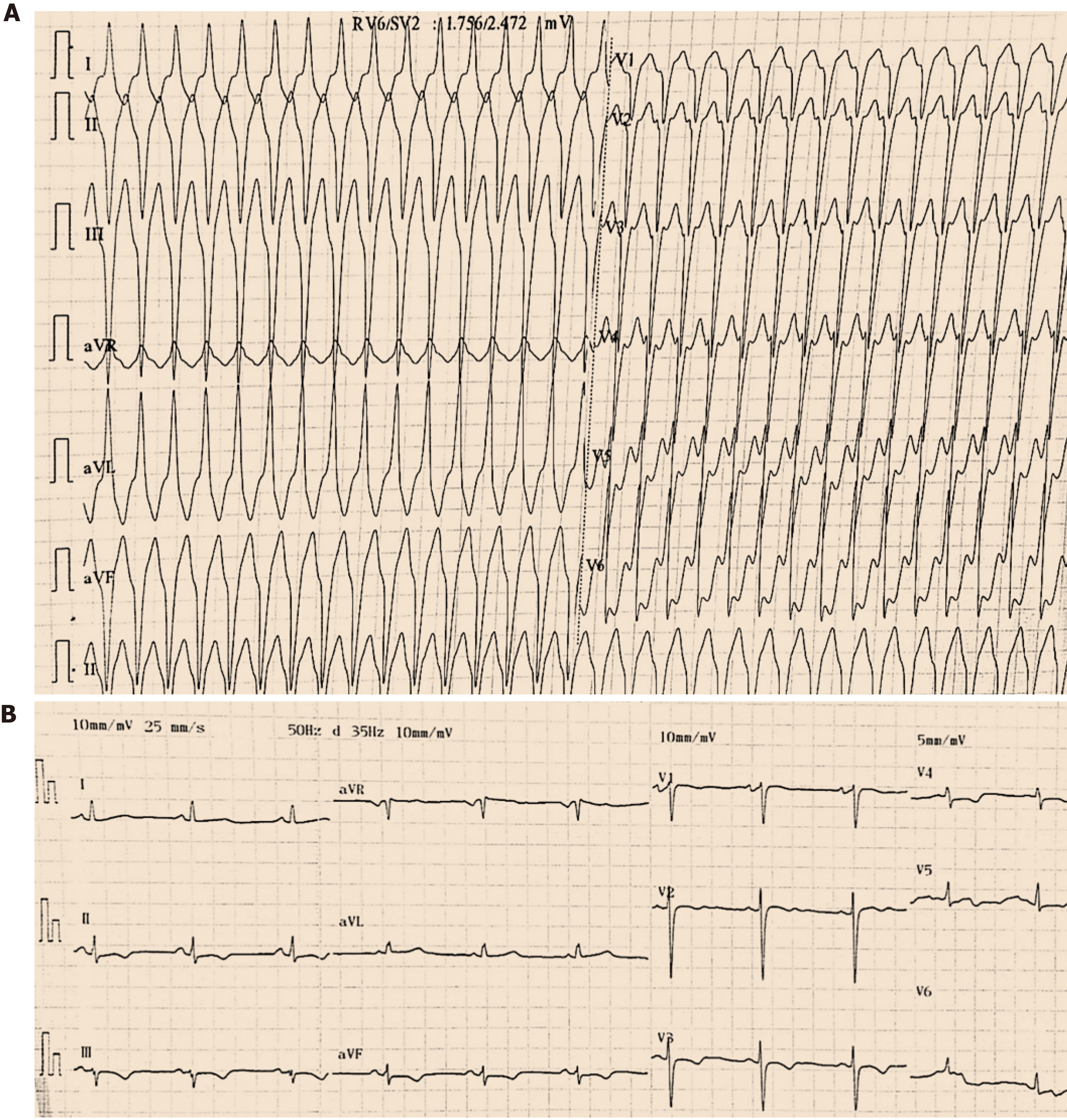
An initial 12-lead ECG at admission demonstrated a regular, wide QRS complex tachycardia at 192 beats per minute of a left bundle-branch block morphology with a broad positive R-wave in lead aVR, an initial R-wave duration greater than 30 ms in lead V2 and an R-to-S interval greater than 100 ms in lead V6. These ECG findings strongly suggested VT (according to the VT morphology criteria in the Brugada algorithm and the initial dominant R-wave in the Vereckei aVR algorithm). The left bundle-branch block morphology and superior axis (positive QRS in lead aVL and negative QRS in leads II, III, and aVF) were consistent with VT originating in the inferior wall of the right ventricle (Figure 1A). The VT was terminated with intravenous propafenone (70 mg). A repeat ECG showed a regular sinus rhythm of 65 beats per minute with negative T waves and a delayed S-wave upstroke (60 ms) from leads V1 to V4 (Figure 1B).
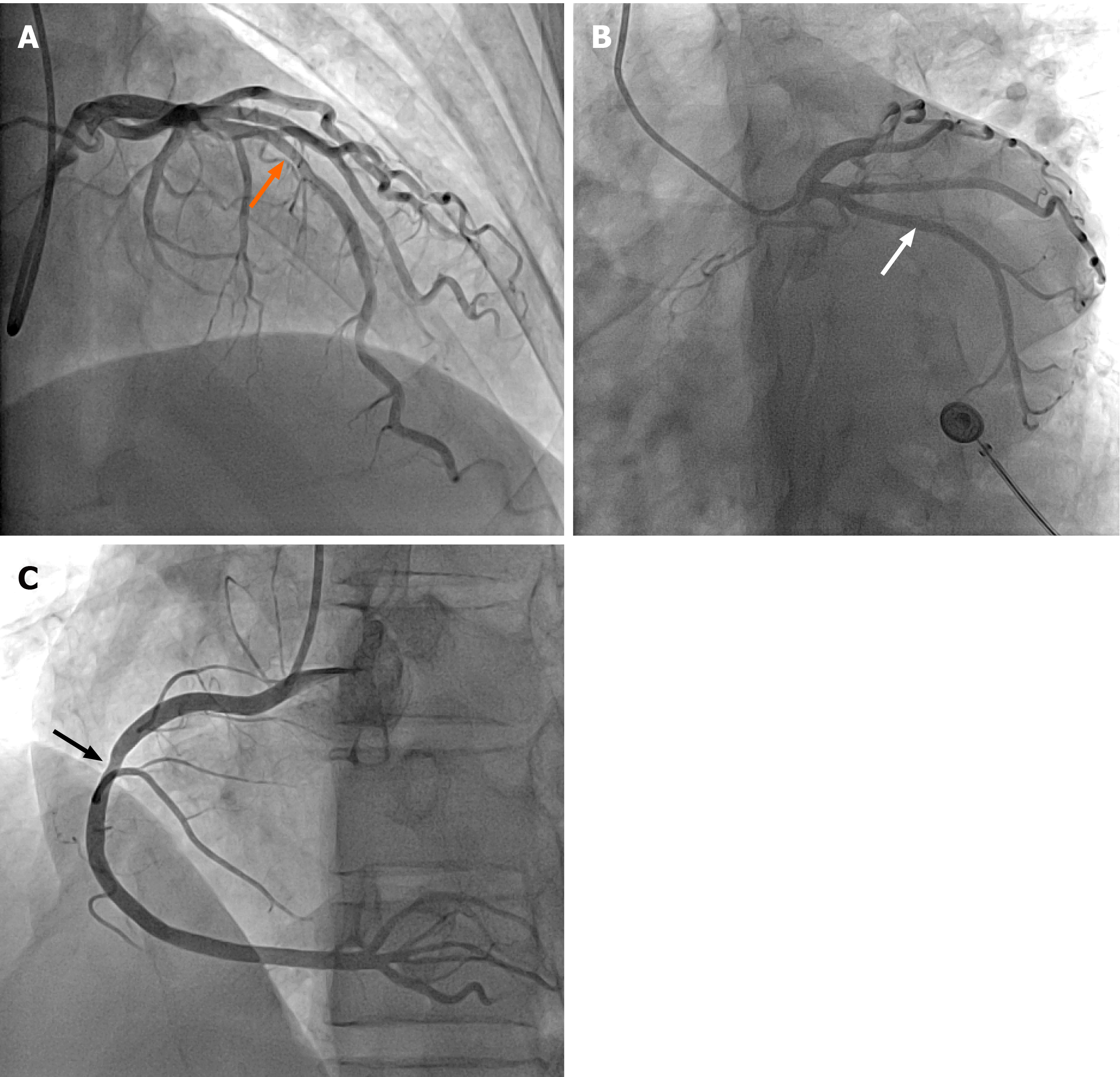
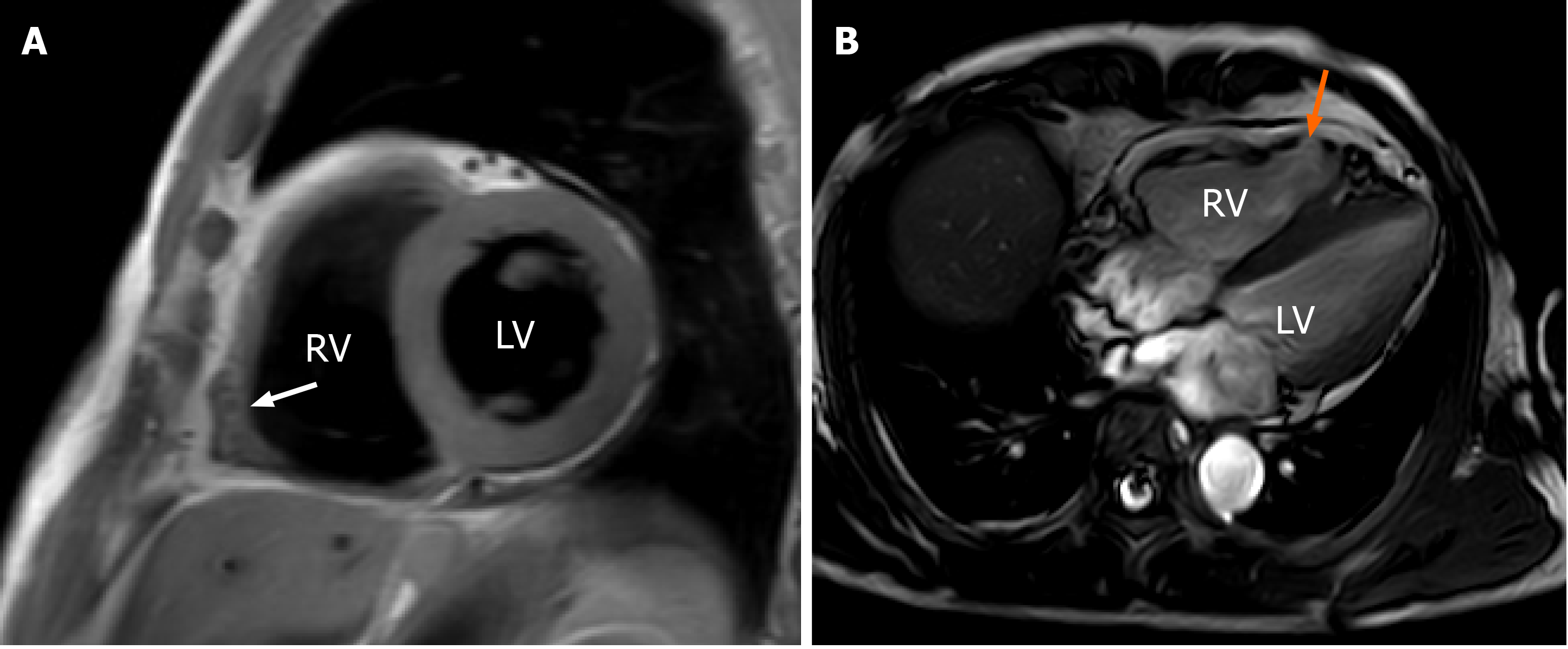
A coronary angiogram revealed mild coronary stenosis (Figure 2), and RV angiography showed regional RV akinesia and aneurysm (Video 1). Cardiac magnetic resonance imaging (CMRI) showed RV free wall thinning, regional akinesia, dilatation, aneurysm and fibrofatty infiltration (RV end-diastolic volume to body surface area 127 mL/m2, RV ejection fraction of 38%), and the left ventricular structure and function were normal (Figure 3).
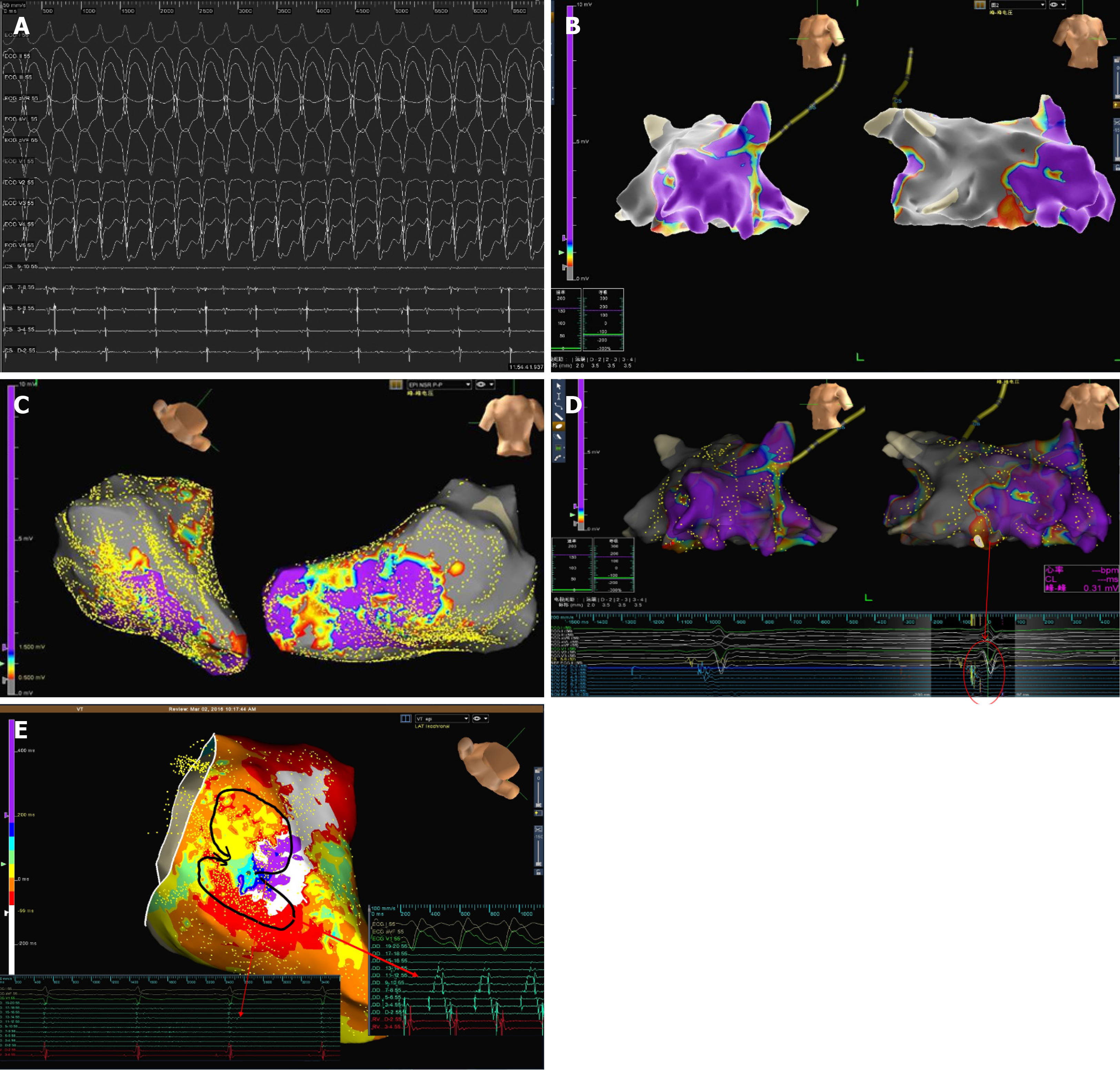
The patient underwent an electrophysiological study, which revealed multiple inducible VTs of a focal mechanism from the right ventricle (Figure 4A). Endocardial and epicardial 3D-electroanatomic voltage mapping demonstrated scar tissue (low-voltage < 0.5 mV) in the anterior wall, free wall and posterior wall of the right ventricle (Figure 4B and 4C). Late potentials were recorded (Figure 4D), and the focal mechanism of VT was marked (Figure 4E).
The diagnosis of ARVC was confirmed.
Combined endocardial and epicardial catheter ablation rendered all VTs noninducible by programmed stimulation with up to four extra stimuli.
The patient was free of complications during hospitalization. Vigorous exercise or exertion was not recommended. At the 4-year follow-up, the patient was asymp
The patient in this case report developed recurrent syncope during exercise. Based on the finding of RV akinesia and a reduced RV ejection fraction on CMRI, along with the presenting arrhythmia and ECG repolarization and depolarization abnormalities, the patient met the revised task force criteria for ARVC (three major and one minor criteria)[5].
ARVC is a rare cardiomyopathy that is mainly determined by genetics, and its pathological feature is fibrofatty substitution of the myocardium, particularly in the right ventricle. In the early stage, structural changes may be absent or slight and are limited to the local area of the right ventricle, usually in the inflow, outflow or apex of the right ventricle. Progression to more diffuse RV disease and left ventricular involvement, with the posterior lateral wall most often involved, can occur. The biventricular variant of ARVC, involving two ventricles, is rare, and its progression is characterized by systolic damage and biventricular dilatation. The clinical features are global congestive heart failure and ventricular arrhythmias originating from either ventricle[6]. Fibrofatty tissue in ARVC develops from the epicardium to the endocardium, resulting in thinning of the ventricular wall, ventricular dilation and aneurysm. Fibrofatty tissue is thought to participate in the occurrence of ventricular arrhythmia by inhibiting intraventricular conduction and scar-related large macro re-entry mechanisms, similar to the situation observed after myocardial infarction[7].
Physical exercise is considered to be a factor that promotes the development and progression of the ARVC phenotype. Exercise can increase the risk of sudden cardiac death in patients with ARVC, which may be related to the aggravation of the mechanical uncoupling of myocardial cells and the resultant malignant ventricular arrhythmia. The damage caused by adhesion between myocardial cells may lead to vulnerability in tissues and organs, which may promote myocardial cell death, especially in the case of mechanical stress, which occurs in endurance exercise. This may increase the age-related penetrance of ARVC desmosome gene carriers, VT risk and heart failure rate[8]. A decrease in exercise after the clinical manifestations of ARVC is independently related to the decrease in ventricular arrhythmia. Regardless of the mutation status and treatment plan of ARVC patients, it should be recommended to restrict exercise, especially for patients with elusive genes, and patients may particularly benefit from a primary prevention implantable cardioverter defibrillator (ICD). In high-risk patients, reducing exercise is unlikely to sufficiently reduce the risk of arrhythmia[9].
ARVC may remain asymptomatic or be associated with multiple symptoms, such as effort-induced syncope, palpitations and dizziness. Sudden cardiac death may be the first manifestation of ARVC, at an average age of 20-40 years[10]. ARVC is more malignant in men than in women, which can be explained by the direct effect of sex hormones on the mechanism of disease phenotype expression[7].
In 2010, the international task force revised the ARVC diagnostic guidelines[5]. Due to the lack of specific diagnostic criteria, the diagnosis of ARVC is still challenging, as there are multiple causes of RV arrhythmias, fairly nonspecific ECG abnormalities, difficulties in imaging the RV structure and function and sometimes confusing gene detection results[7,11]. ECG in patients with ARVC usually shows delayed RV depolarization, manifested by prolonged terminal activation (S wave) duration in leads V1 to V3. Inverted T waves in leads V1 to V3 occurred in up to 87% of patients with ARVC[12]. Epsilon waves may also be recognized in leads V1 to V3 in 30% of ARVC patients, which represent the delayed-activation region in the right ventricle caused by myocardial fibrofatty replacement[13]. However, approximately 30% of patients diagnosed with ARVC do not meet the 2010 ECG criteria[13].
Ventricular arrhythmias are triggered or worsened by adrenergic stimulation, and they include frequent premature ventricular contractions, VT and ventricular fibrillation. Imaging techniques for the diagnosis of ARVC include RV dilation and dysfunction and regional wall motion abnormalities. Because of the anatomical location, load-dependent physiology, complex geometry and challenging acoustic windows of the right ventricle, it is difficult to accurately assess RV structure and function by echocardiography[14]. At the same time, the sensitivity of echocardio
CMRI is the preferred imaging technique for the diagnosis of ARVC because it assesses ventricular structural and functional abnormalities through noninvasive tissue characteristics, especially when late gadolinium enhancement is used, which provides information on fibrofatty myocardial scars[15]. Although endocardial biopsies may be helpful in the differential diagnosis from other cardiomyopathies, they often produce nonspecific findings owing to the patchy nature of the disease[16]. In addition, safety issues limit the use of endocardial biopsies. Although endocardial biopsy is not a routine indication, it should be reserved for patients with a sporadic form of ARVC.
Gene detection has allowed great progress in the diagnosis of ARVC, and it appears to be crucial for excluding ARVC in subjects whose ECG, echocardiography or CMRI cannot provide definite results and for screening relatives of ARVC patients. Sixteen genes have been associated with the ARVC phenotype, and most of them encode components of desmosomes[17]. However, the true prevalence of genetic mutations causing ARVC has not been determined, and the success rate of genotyping is estimated to be approximately 50% in patients who meet the diagnostic criteria for ARVC[18]. Therefore, negative gene testing cannot exclude the possibility of ARVC.
The current treatment of ARVC is palliative, which can only partially relieve symptoms and reduce sudden cardiac death risk. Antiarrhythmic drugs are the first-line treatment for ARVC ventricular arrhythmias with stable hemodynamics. However, there are no prospective and randomized trials on the systematic comparison of antiarrhythmic drug treatment in ARVC. In addition, it is difficult to evaluate the efficacy of specific antiarrhythmic drug treatments because patients with ARVC often develop multiple arrhythmias with the progression of the disease, and antiarrhythmic drugs often change. The currently available data are limited to retrospective analyses, case-control studies and clinical registration. Therefore, the indications and selection of antiarrhythmic drugs are based on empirical methods inferred from other diseases, personal experience, consensus and personal decisions.
Sotalol has been shown to be beneficial for ARVC. Amiodarone alone or in combination with β receptor blockers is considered to be an effective treatment. It is recommended that an ICD can be applied in cardiac arrest survivors or persistent VT patients[14]. An ICD is still the standard of care in patients with ARVC with prior reported VT/syncope. Considering the risks of late ICD system infection, lead failure, inappropriate shocks, vascular obstruction, endocarditis and the high costs, the patient in our case received radiofrequency ablation.
Catheter ablation is an important treatment method for VT in ARVC. Because of the transmural involvement of ARVC in most patients and the early involvement of the epicardium, the initial endocardial ablation-only results were disappointing. The long-term effect of endocardial ablation to prevent recurrence of VT is only 25%-53%[19]. The unsatisfactory results were improved due to the increased understanding of the pathophysiology of ARVC and the application of epicardial ablation. The combination of epicardial and endocardial catheter ablation to map and target all inducible VTs can lead to good long-term outcomes with antiarrhythmic results in most patients without drug treatment. Recent studies have shown that the effectiveness rate of combined epicardial and endocardial ablation to prevent recurrence of VT is 45%-84.6%[20].
Classification of cardiomyopathy based on clinical phenotype is critical. A single test cannot diagnose ARVC. The diagnosis requires comprehensive judgment from morphofunctional, electrophysiological, histological and genetic examinations. In general, ARVC should be suspected in patients with RV arrhythmia, a family history of ARVC or sudden death or abnormal ECG features such as repolarization or depolarization abnormalities (V1 to V3), and further workup, including imaging with multiple modalities (such as CMRI), should be pursued. The combination of epicardial and endocardial catheter ablation can lead to a good outcome.
Manuscript source: Unsolicited manuscript
Specialty type: Medicine, research and experimental
Country/Territory of origin: China
Peer-review report’s scientific quality classification
Grade A (Excellent): A
Grade B (Very good): 0
Grade C (Good): C, C, C
Grade D (Fair): D
Grade E (Poor): 0
P-Reviewer: Al Khader A, Carlier S, Cheng TH, Cismaru G, Esteves M S-Editor: Fan JR L-Editor: Filipodia P-Editor: Xing YX
| 1. | Bawaskar P, Chaurasia A, Nawale J, Nalawade D, Shenoy C. Neoplastic Arrhythmogenic Right Ventricular Cardiomyopathy. Circ Cardiovasc Imaging. 2019;12:e009272. [RCA] [PubMed] [DOI] [Full Text] [Cited by in Crossref: 1] [Cited by in RCA: 1] [Article Influence: 0.2] [Reference Citation Analysis (0)] |
| 2. | Refaat MM, Tang P, Harfouch N, Wojciak J, Kwok PY, Scheinman M. Arrhythmogenic Right Ventricular Cardiomyopathy Caused by a Novel Frameshift Mutation. Card Electrophysiol Clin. 2016;8:217-221. [RCA] [PubMed] [DOI] [Full Text] [Cited by in Crossref: 2] [Cited by in RCA: 2] [Article Influence: 0.2] [Reference Citation Analysis (0)] |
| 3. | Coelho SA, Silva F, Silva J, António N. Athletic Training and Arrhythmogenic Right Ventricular Cardiomyopathy. Int J Sports Med. 2019;40:295-304. [RCA] [PubMed] [DOI] [Full Text] [Cited by in Crossref: 5] [Cited by in RCA: 4] [Article Influence: 0.7] [Reference Citation Analysis (0)] |
| 4. | Prior D, La Gerche A. Exercise and Arrhythmogenic Right Ventricular Cardiomyopathy. Heart Lung Circ. 2020;29:547-555. [RCA] [PubMed] [DOI] [Full Text] [Cited by in Crossref: 22] [Cited by in RCA: 33] [Article Influence: 5.5] [Reference Citation Analysis (0)] |
| 5. | Marcus FI, McKenna WJ, Sherrill D, Basso C, Bauce B, Bluemke DA, Calkins H, Corrado D, Cox MG, Daubert JP, Fontaine G, Gear K, Hauer R, Nava A, Picard MH, Protonotarios N, Saffitz JE, Sanborn DM, Steinberg JS, Tandri H, Thiene G, Towbin JA, Tsatsopoulou A, Wichter T, Zareba W. Diagnosis of arrhythmogenic right ventricular cardiomyopathy/dysplasia: proposed modification of the Task Force Criteria. Eur Heart J. 2010;31:806-814. [RCA] [PubMed] [DOI] [Full Text] [Full Text (PDF)] [Cited by in Crossref: 1156] [Cited by in RCA: 1007] [Article Influence: 67.1] [Reference Citation Analysis (0)] |
| 6. | Bennett RG, Haqqani HM, Berruezo A, Della Bella P, Marchlinski FE, Hsu CJ, Kumar S. Arrhythmogenic Cardiomyopathy in 2018-2019: ARVC/ALVC or Both? Heart Lung Circ. 2019;28:164-177. [RCA] [PubMed] [DOI] [Full Text] [Cited by in Crossref: 37] [Cited by in RCA: 52] [Article Influence: 7.4] [Reference Citation Analysis (0)] |
| 7. | Corrado D, Link MS, Calkins H. Arrhythmogenic Right Ventricular Cardiomyopathy. N Engl J Med. 2017;376:1489-1490. [RCA] [PubMed] [DOI] [Full Text] [Cited by in Crossref: 23] [Cited by in RCA: 40] [Article Influence: 5.0] [Reference Citation Analysis (0)] |
| 8. | Corrado D, Wichter T, Link MS, Hauer R, Marchlinski F, Anastasakis A, Bauce B, Basso C, Brunckhorst C, Tsatsopoulou A, Tandri H, Paul M, Schmied C, Pelliccia A, Duru F, Protonotarios N, Estes NA 3rd, McKenna WJ, Thiene G, Marcus FI, Calkins H. Treatment of arrhythmogenic right ventricular cardiomyopathy/dysplasia: an international task force consensus statement. Eur Heart J. 2015;36:3227-3237. [RCA] [PubMed] [DOI] [Full Text] [Full Text (PDF)] [Cited by in Crossref: 24] [Cited by in RCA: 80] [Article Influence: 8.0] [Reference Citation Analysis (0)] |
| 9. | Wang W, Orgeron G, Tichnell C, Murray B, Crosson J, Monfredi O, Cadrin-Tourigny J, Tandri H, Calkins H, James CA. Impact of Exercise Restriction on Arrhythmic Risk Among Patients With Arrhythmogenic Right Ventricular Cardiomyopathy. J Am Heart Assoc. 2018;7. [RCA] [PubMed] [DOI] [Full Text] [Full Text (PDF)] [Cited by in Crossref: 51] [Cited by in RCA: 59] [Article Influence: 8.4] [Reference Citation Analysis (0)] |
| 10. | Adesina GO, Hall SA, Mendez JC, Joseph SM, Gottlieb RL, Kale PP, Bindra AS. Arrhythmogenic Right Ventricular Dysplasia: An Under-recognized Form of Inherited Cardiomyopathy. Rev Cardiovasc Med. 2017;18:37-43. [PubMed] |
| 11. | Lopez-Ayala JM, Oliva-Sandoval MJ, Sanchez-Muñoz JJ, Gimeno JR. Arrhythmogenic right ventricular cardiomyopathy. Lancet. 2015;385:662. [RCA] [PubMed] [DOI] [Full Text] [Cited by in Crossref: 4] [Cited by in RCA: 4] [Article Influence: 0.4] [Reference Citation Analysis (0)] |
| 12. | Quarta G, Muir A, Pantazis A, Syrris P, Gehmlich K, Garcia-Pavia P, Ward D, Sen-Chowdhry S, Elliott PM, McKenna WJ. Familial evaluation in arrhythmogenic right ventricular cardiomyopathy: impact of genetics and revised task force criteria. Circulation. 2011;123:2701-2709. [RCA] [PubMed] [DOI] [Full Text] [Cited by in Crossref: 181] [Cited by in RCA: 194] [Article Influence: 13.9] [Reference Citation Analysis (0)] |
| 13. | Gandjbakhch E, Redheuil A, Pousset F, Charron P, Frank R. Clinical Diagnosis, Imaging, and Genetics of Arrhythmogenic Right Ventricular Cardiomyopathy/Dysplasia: JACC State-of-the-Art Review. J Am Coll Cardiol. 2018;72:784-804. [RCA] [PubMed] [DOI] [Full Text] [Cited by in Crossref: 126] [Cited by in RCA: 193] [Article Influence: 32.2] [Reference Citation Analysis (0)] |
| 14. | Hamilton-Craig C, McGavigan A, Semsarian C, Martin A, Atherton J, Stanton T, La Gerche A, Taylor AJ, Haqqani H. The Cardiac Society of Australia and New Zealand Position Statement on the Diagnosis and Management of Arrhythmogenic Right Ventricular Cardiomyopathy (2019 Update). Heart Lung Circ. 2020;29:40-48. [RCA] [PubMed] [DOI] [Full Text] [Cited by in Crossref: 2] [Cited by in RCA: 2] [Article Influence: 0.3] [Reference Citation Analysis (0)] |
| 15. | Steinmetz M, Krause U, Lauerer P, Konietschke F, Aguayo R, Ritter CO, Schuster A, Lotz J, Paul T, Staab W. Diagnosing ARVC in Pediatric Patients Applying the Revised Task Force Criteria: Importance of Imaging, 12-Lead ECG, and Genetics. Pediatr Cardiol. 2018;39:1156-1164. [RCA] [PubMed] [DOI] [Full Text] [Cited by in Crossref: 17] [Cited by in RCA: 21] [Article Influence: 3.0] [Reference Citation Analysis (0)] |
| 16. | Brandimarte F, Battagliese A, Pirillo SP, Mallus MT, Manfredi RM, Carreras G. A case of arrhythmogenic right ventricular cardiomyopathy with biventricular involvement. Monaldi Arch Chest Dis. 2019;89. [RCA] [PubMed] [DOI] [Full Text] [Cited by in Crossref: 1] [Cited by in RCA: 1] [Article Influence: 0.2] [Reference Citation Analysis (0)] |
| 17. | Awad MM, Calkins H, Judge DP. Mechanisms of disease: molecular genetics of arrhythmogenic right ventricular dysplasia/cardiomyopathy. Nat Clin Pract Cardiovasc Med. 2008;5:258-267. [RCA] [PubMed] [DOI] [Full Text] [Full Text (PDF)] [Cited by in Crossref: 203] [Cited by in RCA: 181] [Article Influence: 10.6] [Reference Citation Analysis (0)] |
| 18. | Bhonsale A, Groeneweg JA, James CA, Dooijes D, Tichnell C, Jongbloed JD, Murray B, te Riele AS, van den Berg MP, Bikker H, Atsma DE, de Groot NM, Houweling AC, van der Heijden JF, Russell SD, Doevendans PA, van Veen TA, Tandri H, Wilde AA, Judge DP, van Tintelen JP, Calkins H, Hauer RN. Impact of genotype on clinical course in arrhythmogenic right ventricular dysplasia/cardiomyopathy-associated mutation carriers. Eur Heart J. 2015;36:847-855. [RCA] [PubMed] [DOI] [Full Text] [Cited by in Crossref: 268] [Cited by in RCA: 319] [Article Influence: 31.9] [Reference Citation Analysis (0)] |
| 19. | Dalal D, Jain R, Tandri H, Dong J, Eid SM, Prakasa K, Tichnell C, James C, Abraham T, Russell SD, Sinha S, Judge DP, Bluemke DA, Marine JE, Calkins H. Long-term efficacy of catheter ablation of ventricular tachycardia in patients with arrhythmogenic right ventricular dysplasia/cardiomyopathy. J Am Coll Cardiol. 2007;50:432-440. [RCA] [PubMed] [DOI] [Full Text] [Cited by in Crossref: 192] [Cited by in RCA: 164] [Article Influence: 9.1] [Reference Citation Analysis (0)] |
| 20. | Chung FP, Lin CY, Lin YJ, Chang SL, Lo LW, Hu YF, Tuan TC, Chao TF, Liao JN, Chang TY, Chen SA. Catheter Ablation of Ventricular Tachycardia in Arrhythmogenic Right Ventricular Dysplasia/Cardiomyopathy. Korean Circ J. 2018;48:890-905. [RCA] [PubMed] [DOI] [Full Text] [Full Text (PDF)] [Cited by in Crossref: 2] [Cited by in RCA: 2] [Article Influence: 0.3] [Reference Citation Analysis (0)] |


