Published online Jun 6, 2021. doi: 10.12998/wjcc.v9.i16.3908
Peer-review started: August 4, 2020
First decision: December 14, 2020
Revised: January 13, 2021
Accepted: April 20, 2021
Article in press: April 20, 2021
Published online: June 6, 2021
Processing time: 283 Days and 1.8 Hours
A snapping wrist is a rare symptom that results from the sudden impingement of one anatomic structure against another, subsequently causing a sudden movement only during wrist movement.
A 30-year-old woman with a history of right wrist contusion reported right wrist snapping after overuse. The snapping became symptomatic after moving heavy objects. The pain persisted even when she received 1 mo of conservative treatment. Physical examination showed painful wrist snapping during wrist radioulnar motion and thumb abduction-adduction. Radiography demonstrated bone overgrowth over the radial styloid process. Sonography disclosed a tendon jumping over a bony prominence in the first compartment during wrist motion. Magnetic resonance imaging revealed no anomalous tendon nor tumorlike lesion. Under the wide-awake local anesthesia no tourniquet (WALANT) technique, the lesion was identified in the first extensor compartment. The patient received stepwise extensor retinaculum release, synovectomy, and bone spur removal. By 6th week, the patient was completely free of pain and unable to snap her wrist. She started working 7 wk after the surgery. One year after the surgery, the wrist snap was not recurrent.
Careful physical examination and dynamic sonography may confirm the diagnosis of a snapping wrist. With the WALANT technique, the lesion could be identified under direct vision, and we could take stepwise interventions according to intraoperative presentations.
Core Tip: Wrist snapping is a rare symptom in clinical scenario. We reported a young lady with asymptomatic wrist snapping that became painful after long term overuse. This study offered dynamic sonographic images and intraoperative pictures to identify the pathogen of the snapping wrist, which was not reported by previous research. Besides, we presented stepwise interventions in the surgery and showed the advantage of the wide-awake local anesthesia no tourniquet technique in treating our patient.
- Citation: Hu CJ, Chow PC, Tzeng IS. Snapping wrist due to bony prominence and tenosynovitis of the first extensor compartment: A case report. World J Clin Cases 2021; 9(16): 3908-3913
- URL: https://www.wjgnet.com/2307-8960/full/v9/i16/3908.htm
- DOI: https://dx.doi.org/10.12998/wjcc.v9.i16.3908
Snapping syndromes result from the sudden impingement of one anatomic structure against another, subsequently causing a sudden movement. Though clunking, clicking, locking, and triggering are interchangeable terms in many anatomic regions, some authors suggest that trigger wrist and snapping wrist are different descriptions for patients with various presentations[1]. Trigger wrist mainly refers to patients with trigger finger at the volar wrist that is often associated with pain and carpal tunnel syndrome. The clicking sound can be produced by the movement of the wrist or fingers[2-4]. On the other hand, a snapping wrist occurs only during wrist movement. The location of clicking varies from the dorsal wrist compartment and intercarpal joint[5] to the volar wrist. Neurologic symptoms may not present in volar wrist snapping[6].
We report a rare case of dorso-radial wrist snapping due to bony prominence and tenosynovitis of the first extensor compartment. To the best of our knowledge, this is the first report on the pathogenesis of snapping wrist caused by this condition.
A 30-year-old right-handed female Filipino migrant worker visited our clinic with a 7-year history of right wrist snapping.
The patient had right painful wrist snapping when she moved heavy objects during work 2 years ago. The symptoms exacerbated in recent months and did not improve after use of analgesics and a 4-wk rest.
She had a wrist pain episode after a fall with an outstretched hand 7 years ago. She completely recovered after a 4-wk conservative treatment. She found her wrist snapped while cooking about 10 mo after the fall episode. However, neither pain nor neurologic symptoms were provoked when the snap occurred.
She did not smoke and reported no specific family history.
Physical examination showed snapping at the radial wrist during wrist radioulnar motion and thumb abduction-adduction. The pain was induced only with a snap and localized to the first dorsal compartment, just 1 cm proximal to the radial styloid process. No tenderness around the radial styloid process, no limitation of right wrist range of motion, no loss of grip strength, no alteration in sensation, and no wrist instability were noted. Except the moment the snap occurred, no pain was reported during the Finkelstein test.
Serum laboratory data, including complete blood count with differential and rheumatoid factor, were within normal limits.
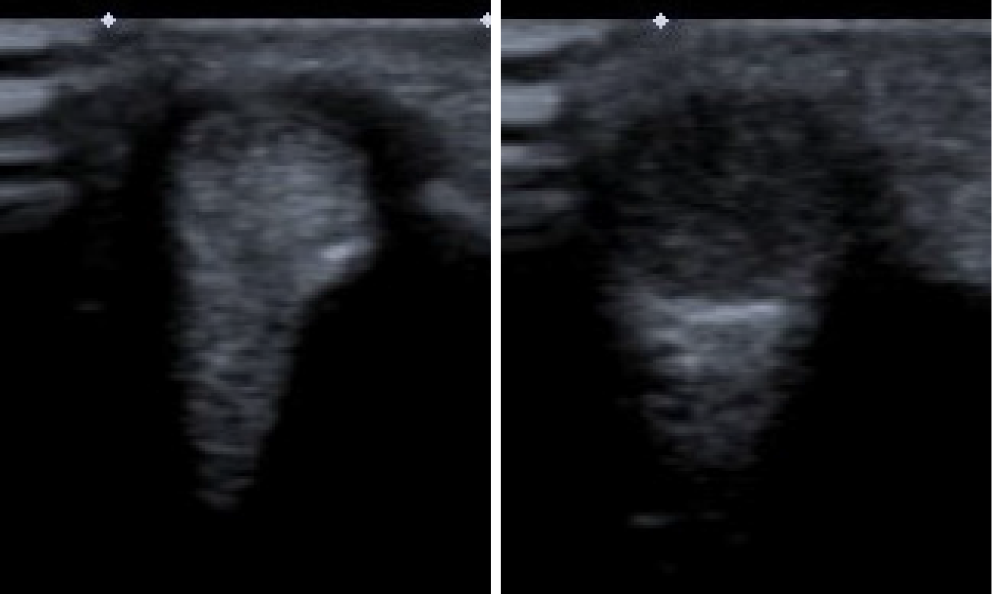
Plain film of the right wrist showed bony overgrowth over the radial styloid process. Dynamic sonography disclosed a tendon jumping over a bony prominence in the first compartment during wrist motion (Figure 1). Magnetic resonance imaging (MRI) revealed no anomalous tendon nor tumorlike lesion.
The final diagnosis was right snapping wrist related to bony prominence and tenosynovitis of the first extensor compartment.
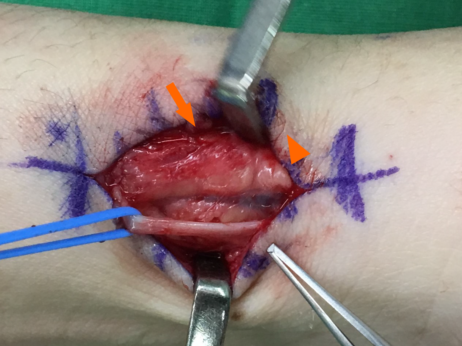
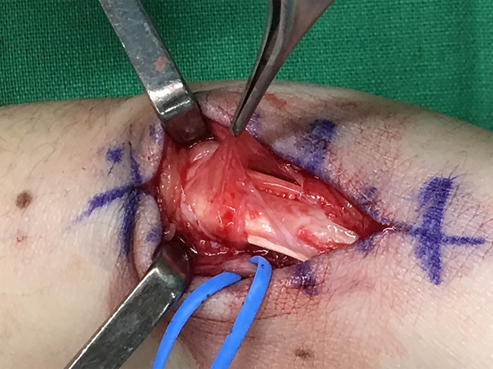
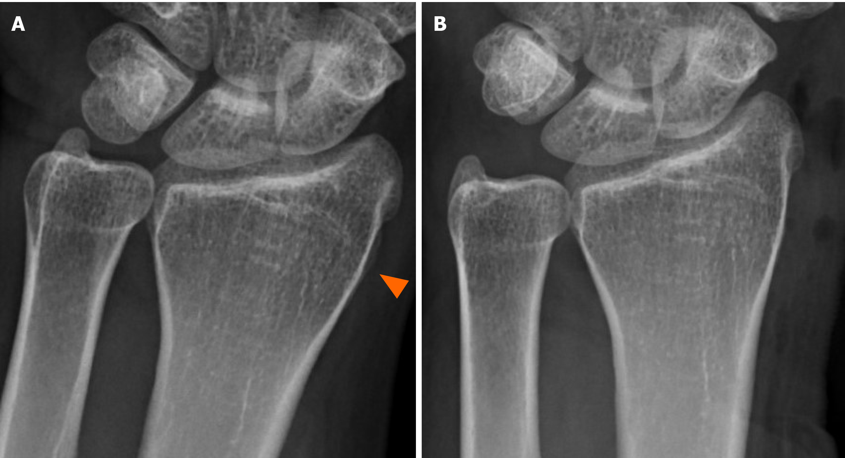
Under the wide-awake local anesthesia no tourniquet (WALANT) technique, the snapping phenomenon was identified as the beneath enlarged tendon gliding out of the distal extensor retinaculum, forming a soft tissue ball (Figure 2). The retinaculum was released at the first extensor dorsal compartment, and the soft tissue ball was recognized as a proliferated synovium from the abductor pollicis longus (APL). After synovectomy, two slips of APL were identified (Figure 3). Although the wrist snap subsided after retinaculum release, we still found uneven tendons gliding over a bony prominence during wrist motion. Therefore, we subperiosteally elevated the first extensor compartment and removed the bony spur to deepen the gliding groove (Figure 4). Smooth tendon glide was confirmed through wrist radioulnar motion and thumb abduction-adduction.
By 6th week, the patient was completely free of pain and unable to snap her wrist. She started working 7 wk after the surgery. One year after the surgery, the wrist snap was not recurrent.
The etiology of a snapping wrist could be classified into the intra- and extracapsular regions[7]. In intracapsular pathology, carpal instabilities between or within the carpal rows are the most common causes. In addition, loose bodies[8], thickening of the dorsal wrist capsule, and hypertrophic scarring of the carpal ligament[7] could lead to the snapping phenomenon. For extracapsular lesions, tumors or masses accounted for most of the etiology. These lesions usually arise from the muscle[3,9], tendons[10], or tendon sheath[4,11] and may be a result of a ruptured muscle[12], anatomic varia
In our case, the wrist snapping was due to the extracapsular synovial ball from one slip of the APL tendon. It is supposed that long-standing overuse induced synovial proliferation. We released the retinaculum and excised the synovial ball to prevent the snap phenomenon. The bony prominence of the distal radius was removed after evaluating the tendon slide during surgery. Though multiple tendon slips in the first extensor compartment are an etiology of a snapping wrist[10], we did not excise any slip of the APL. A previous study was an extreme case with multiple accessory APL and extensor pollicis brevis that caused extracapsular mass effect. However, approximately 80% of normal people experience multiple slips of APL, and these slips alone are not risk factors for tenosynovitis of the first extensor compartment[13]. Synovectomy and ostectomy may be sufficient for our patient.
Careful physical examination and dynamic sonography are key to confirm the diagnosis of a snapping wrist. MRI is an assisted tool to clarify possible lesions. With the WALANT technique, the lesion could be identified under direct vision, and we could take stepwise interventions according to intraoperative presentations.
Manuscript source: Unsolicited manuscript
Specialty type: Medicine, research and experimental
Country/Territory of origin: Taiwan
Peer-review report’s scientific quality classification
Grade A (Excellent): 0
Grade B (Very good): B
Grade C (Good): 0
Grade D (Fair): 0
Grade E (Poor): 0
P-Reviewer: Zhu YF S-Editor: Gao CC L-Editor: A P-Editor: Xing YX
| 1. | Park IJ, Lee YM, Kim HM, Lee JY, Roh YT, Park CK, Kang SH. Multiple etiologies of trigger wrist. J Plast Reconstr Aesthet Surg. 2016;69:335-340. [RCA] [PubMed] [DOI] [Full Text] [Cited by in Crossref: 5] [Cited by in RCA: 8] [Article Influence: 0.8] [Reference Citation Analysis (0)] |
| 2. | Sonoda H, Takasita M, Taira H, Higashi T, Tsumura H. Carpal tunnel syndrome and trigger wrist caused by a lipoma arising from flexor tenosynovium: a case report. J Hand Surg Am. 2002;27:1056-1058. [RCA] [PubMed] [DOI] [Full Text] [Cited by in Crossref: 24] [Cited by in RCA: 28] [Article Influence: 1.2] [Reference Citation Analysis (0)] |
| 3. | Shimizu A, Ikeda M, Kobayashi Y, Saito I, Mochida J. Carpal Tunnel Syndrome with Wrist Trigger Caused by Hypertrophied Lumbrical Muscle and Tenosynovitis. Case Rep Orthop. 2015;2015:705237. [RCA] [PubMed] [DOI] [Full Text] [Full Text (PDF)] [Cited by in Crossref: 5] [Cited by in RCA: 8] [Article Influence: 0.8] [Reference Citation Analysis (0)] |
| 4. | Chalmers RL, Mandalia M, Contreras R, Schreuder F. Acute trigger wrist and carpal tunnel syndrome due to giant-cell tumour of the flexor sheath. J Plast Reconstr Aesthet Surg. 2008;61:1557. [RCA] [PubMed] [DOI] [Full Text] [Cited by in Crossref: 6] [Cited by in RCA: 5] [Article Influence: 0.3] [Reference Citation Analysis (0)] |
| 5. | Chinder PS, Kamath BJ, Hegde D, Rai M. Snapping wrist due to lunate malformation. Indian J Plast Surg. 2012;45:581-583. [RCA] [PubMed] [DOI] [Full Text] [Cited by in Crossref: 2] [Cited by in RCA: 2] [Article Influence: 0.2] [Reference Citation Analysis (0)] |
| 6. | Itsubo T, Uchiyama S, Takahara K, Nakagawa H, Kamimura M, Miyasaka T. Snapping wrist after surgery for carpal tunnel syndrome and trigger digit: a case report. J Hand Surg Am. 2004;29:384-386. [RCA] [PubMed] [DOI] [Full Text] [Cited by in Crossref: 15] [Cited by in RCA: 15] [Article Influence: 0.7] [Reference Citation Analysis (0)] |
| 7. | Swann RP, Noureldin M, Kakar S. Dorsal Radiotriquetral Ligament Snapping Wrist Syndrome - A Novel Presentation and Review of Literature: Case Report. J Hand Surg Am 2016; 41: 344-7. e2. [RCA] [PubMed] [DOI] [Full Text] [Cited by in Crossref: 5] [Cited by in RCA: 6] [Article Influence: 0.7] [Reference Citation Analysis (0)] |
| 8. | Zachee B, DeSmet L, Fabry G. A snapping wrist due to a loose body. Arthroscopic diagnosis and treatment. Arthroscopy. 1993;9:117-118. [RCA] [PubMed] [DOI] [Full Text] [Cited by in Crossref: 18] [Cited by in RCA: 13] [Article Influence: 0.4] [Reference Citation Analysis (0)] |
| 9. | Baker J, Gonzalez MH. Snapping wrist due to an anomalous extensor indicis proprius: a case report. Hand (N Y). 2008;3:363-365. [RCA] [PubMed] [DOI] [Full Text] [Cited by in Crossref: 8] [Cited by in RCA: 9] [Article Influence: 0.5] [Reference Citation Analysis (0)] |
| 10. | Subramaniyam SD, Purushothaman R, Zacharia B. Snapping wrist due to multiple accessory tendon of first extensor compartment. Int J Surg Case Rep. 2018;42:182-186. [RCA] [PubMed] [DOI] [Full Text] [Full Text (PDF)] [Cited by in Crossref: 1] [Cited by in RCA: 1] [Article Influence: 0.1] [Reference Citation Analysis (0)] |
| 11. | Eibel P. Trigger Wrist with Intermittent Carpal Tunnel Syndrome: A Hitherto Undescribed Entity with Report of a Case. Can Med Assoc J. 1961;84:602-605. [PubMed] |
| 12. | Yamazaki H, Uchiyama S, Kato H. Snapping wrist caused by tenosynovitis of the extensor carpi radialis longus tendon subsequent to subcutaneous muscle rupture in the forearm: case report. J Hand Surg Am. 2010;35:1964-1967. [RCA] [PubMed] [DOI] [Full Text] [Cited by in Crossref: 4] [Cited by in RCA: 5] [Article Influence: 0.3] [Reference Citation Analysis (0)] |
| 13. | Lee ZH, Stranix JT, Anzai L, Sharma S. Surgical anatomy of the first extensor compartment: A systematic review and comparison of normal cadavers vs. De Quervain syndrome patients. J Plast Reconstr Aesthet Surg. 2017;70:127-131. [RCA] [PubMed] [DOI] [Full Text] [Cited by in Crossref: 26] [Cited by in RCA: 44] [Article Influence: 4.9] [Reference Citation Analysis (0)] |


