Published online Apr 26, 2021. doi: 10.12998/wjcc.v9.i12.2868
Peer-review started: November 29, 2020
First decision: December 24, 2020
Revised: December 26, 2020
Accepted: January 27, 2021
Article in press: January 27, 2021
Published online: April 26, 2021
Processing time: 136 Days and 18.7 Hours
‘Splenosis’ is defined as the autotransplantation of splenic tissue following trauma or surgery, usually in the form of intraperitoneal nodules. The proliferation of imaging techniques has resulted in increased unexpected discoveries of splenosis nodules, and achieving a differential diagnosis can be challenging. Nuclear medicine studies have been playing an increasingly important role in this process, but the clinical significance of asymptomatic nodules remains uncertain.
We present a case of pelvic splenosis in a 73-year-old man diagnosed 56 years after a splenectomy during a computed tomography (CT) follow-up for B-cell lymphoma, presenting intense contrast enhancement of an 18 mm nodule in the right recto-vesical space. 18F-fluorodeoxyglucose demonstrated weak metabolic activity. Since histological diagnosis was deemed necessary, the nodule was easily removed with robotically assisted laparoscopy, together with another 6 mm left a paracolic lesion. The latter was previously undiagnosed but retrospectively visible on the CT scan.
In a patient requiring differential diagnosis of splenosis nodules from lymphoma recurrence, the robotic approach provided a safe en bloc removal with short hospitalization. The Da Vinci Xi robot was particularly helpful because its optics can be introduced from all ports, facilitating visualization and lysis of multiple intra-abdominal adhesions.
Core Tip: Splenosis consists in the autotrasplantation of splenic tissue caused by trauma or surgery, usually in the form of asymptomatic intraperitoneal nodules. They usually present intense enhancement at computed tomography scan and nuclear medicine studies have an increasing role in diagnosis. When histological diagnosis is deemed necessary robotically assisted laparoscopic surgery can provide safe en bloc removal with minimal invasiveness.
- Citation: Tognarelli A, Faggioni L, Erba AP, Faviana P, Durante J, Manassero F, Selli C. Robotically assisted removal of pelvic splenosis fifty-six years after splenectomy: A case report. World J Clin Cases 2021; 9(12): 2868-2873
- URL: https://www.wjgnet.com/2307-8960/full/v9/i12/2868.htm
- DOI: https://dx.doi.org/10.12998/wjcc.v9.i12.2868
The term ‘splenosis’ was created 80 years ago to describe the heterotopic autotransplantation of splenic tissue following rupture of the spleen, generally caused by trauma[1]. In general, this condition appears in the form of nodules within the peritoneal cavity with a pattern simulating metastatic spread[2]. Implants have also been discovered within the kidneys, liver, pancreas, brain, chest and subcutaneous fat. Beyond this, a recent nuclear medicine study revealed the presence of residual functional splenic tissue in 34.8% of patients who underwent splenectomy for trauma[3]. Notably, this tissue may provide ongoing immunological protection. The clinical significance of this finding is still unclear because the patients were asymptomatic, but it highlights the association between a history of splenectomy after trauma and the incidental discovery of lesions due to the further development of imaging techniques. Therefore, splenosis should be considered in the differential diagnosis of a mass discovered years after splenic trauma or surgery.
A 73-year-old man was admitted to our urology unit for the treatment of a mass arising in his right recto-vesical space incidentally discovered during a computed tomography (CT) scan.
The patient was asymptomatic.
Past history revealed an appendectomy at age 12; a splenectomy at age 16 due to a motorcycle accident that caused abdominal trauma (56 years earlier); the transurethral resection of a low-grade, non-muscle-invasive bladder tumour at age 71; and B-cell lymphoma of the marginal zone diagnosed with an osteomedullary biopsy at age 72, which was under active surveillance at the time.
The patient had no personal history of illness, and the family history was negative for inherent disease.
The patient was stable, vital signs were normal and the only abnormal finding was the presence of a midline xyphopubic surgical scar with a transverse extension on the left upper quadrant.
No abnormalities were found on routine pre-surgical laboratory examinations, including a complete blood cell count and coagulation profile.
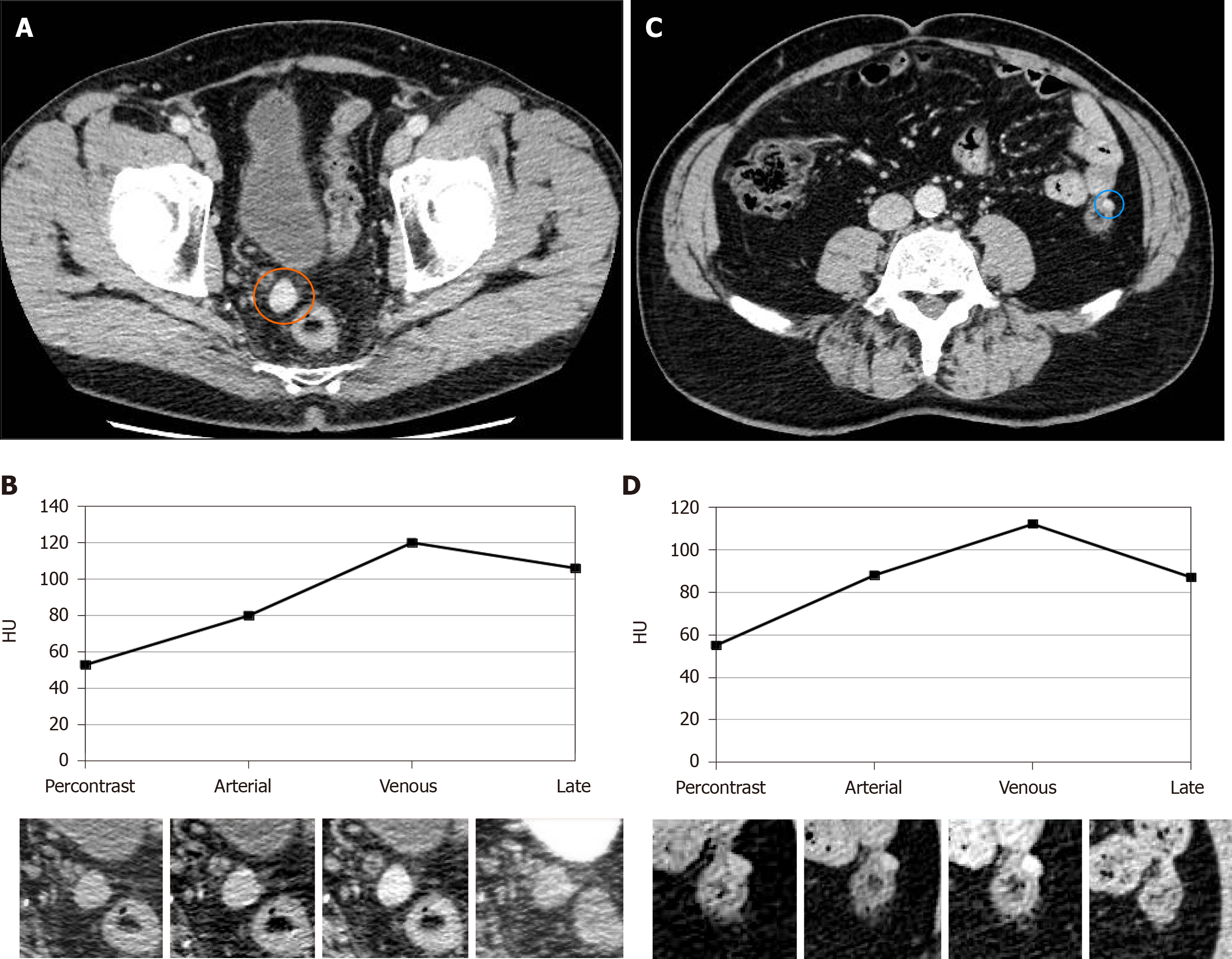
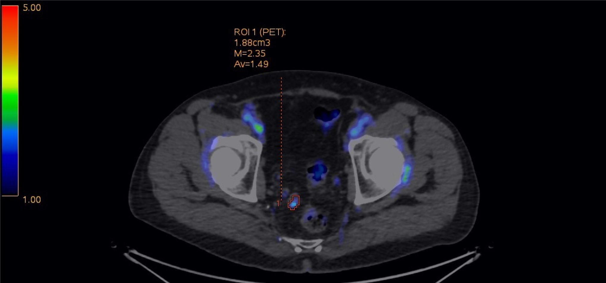
Following the diagnosis of lymphoma one year earlier, a staging contrast-enhanced CT examination revealed an 18 mm rounded nodule in the right recto-vesical space with intense contrast enhancement, tentatively diagnosed as a lymph node. A follow-up CT scan six months later showed that the lesion had remained stable in size (Figure 1A and B). Further evaluation suggested by the haematologist consisted of a 18F-fluorodeoxyglucose (18F-FDG) positron emission tomography (PET)/CT scan, revealing weak metabolic activity (Standardized Uptake Value: 2.35) (Figure 2).
A multidisciplinary oncological evaluation suggested the need for further histological characterization of the lesion presenting enhancement during the CT scan to rule out the possibility of lymphoma.
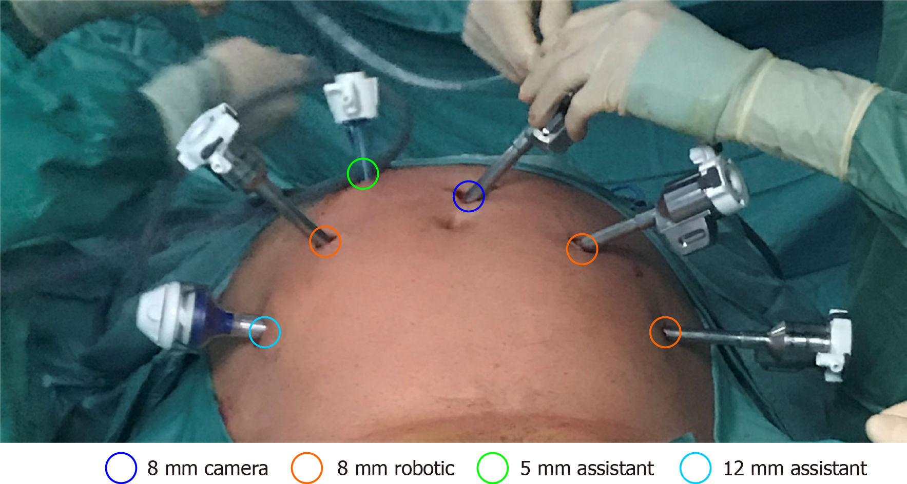
Minimally invasive surgery was preferred through biopsy due to the location of the nodule and the extremely long period of time elapsed since the splenic trauma. A robotic approach was adopted with the standard placement of the six ports as in transperitoneal robotically assisted radical prostatectomy using a Da Vinci XI (Intuitive Surgical Inc, Sunnyvale, CA, United States) (Figure 3). The 8 mm camera port was placed with the Hasson open technique on the midline 3 cm above the umbilicus; the two 8 mm ports for the robotic arms laterally to the rectus muscles at 10 cm from the camera, 2 cm below the umbilicus, the third 8 mm robotic port on the left side 10 cm laterally, along the same line; the 5 mm assistant port was placed in the right upper quadrant (on the mid-clavicular line 5 cm below the costal margin) and the 12 mm assistant port in the right inferior quadrant (on the anterior axillary line 4 cm below the 5 mm assistant port). The Xi robot was then docked on the left side of the patient. Lysis of multiple small bowel adhesions due to previous surgery was necessary, requiring the shifting of the 8 mm optics between different ports.
A purple rounded nodule on the right side of the Douglas pouch was identified, fixed to the retrovesical peritoneum and extending into the pararectal fat. It was easily removed en bloc using monopolar cautery, placed inside a small retrieval bag and extracted at the end of the procedure without touching other tissues. Another 6 mm nodule of similar appearance adherent to the peritoneum overlying the sigmoid colon was also removed. Retrospective evaluation of CT images demonstrated intense contrast enhancement of this smaller lesion as well (Figure 1C and D).
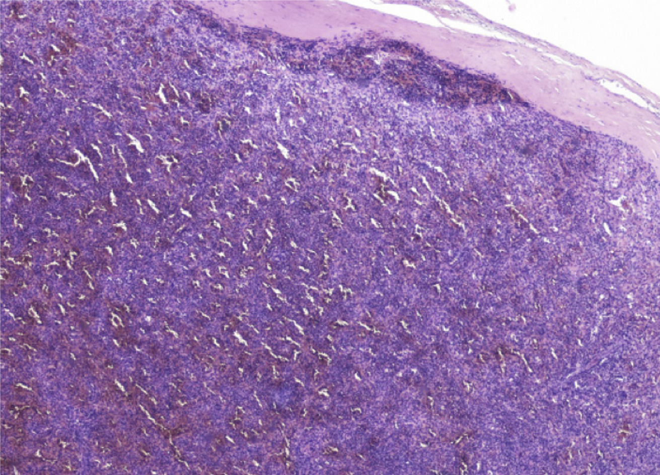
The operation was completed without complications, the postoperative course was uneventful and the patient was discharged on the second postoperative day. On gross inspection, the lesions were purple in colour, rounded and of medium consistency. The histopathological diagnosis of both nodules was splenic tissue (Figure 4).
The patient remained asymptomatic for one year without further nodules appearing during the control CT scan performed for the follow-up to eliminate the possibility of lymphoma recurrences.
The present case presented several interesting aspects, such as the very long interval (56 years) between the splenectomy and the detection of splenosis, the potentially misleading use of imaging techniques, and the effective and minimally invasive treatment with robotically assisted laparoscopy.
During CT, splenosis is characterized by the presence of multiple, typically rounded nodules ranging from a few millimetres to several centimetres in diameter, with densities and contrast enhancement patterns similar to those of the spleen, or the expected density and contrast enhancement of the spleen if splenectomy has been performed[4]. As with a normal spleen, these nodules show a mottled contrast enhancement pattern during the arterial and early portal venous phases. This pattern is due to the existence of different flow rates between the open and closed splenic circulation, and it becomes homogeneous in the late venous and equilibrium phases[5]. The spleen-like contrast enhancement behaviour of splenosis nodules during multiphase contrast-enhanced CT is pathognomonic. However, this behaviour may not be evident in smaller or less vascularized nodules or without the use of a dedicated CT protocol. Moreover, diagnosis can be difficult in post-splenectomy patients due to the absence of the spleen as reference tissue, especially if splenosis nodules are outside the peritoneal cavity or located in the liver, where they may mimic focal primary or secondary liver disease and represent diagnostic challenges[6].
Nuclear medicine techniques have a crucial function in the differential diagnosis of splenosis because residual splenic tissue presents an uptake of heat-denatured erythrocytes labelled with 99mTC at scintigraphy and/or single-photon emission tomography/CT[7], or 99mTC-labeled sulphur colloid. Uptake of specific radiopharmace-uticals such as 68Ga-radiolabelled prostate-specific membrane antigen peptide[8] and 68Ga-labelled somatostatin analogues[9] has been described in patients with splenosis undergoing PET/CT. Furthermore, 18F-FDG activity accumulation can be variable[10].
Surgical treatment has been generally reserved for cases of splenosis presenting with pain, bleeding or obstructive complications. In addition, the use of minimally invasive techniques such as laparoscopy and robotics is ideal for providing safe removal with limited hospitalization[11]. Finally, laparoscopy provides the opportunity for a definitive histological diagnosis.
Urologists are particularly skilled in the robotic treatment of masses arising in the male pelvis. This is because radical robotic prostatectomy with a transperitoneal approach previews the opening of the peritoneum of the Douglas cul-de-sac to isolate the seminal vesicles and to detach the posterior aspect of the prostate from the rectum. The use of the Da Vinci XI robot in the present case with multiple bowel adhesions secondary to previous surgery proved to be particularly helpful. This was the case because the 8 mm optics could be introduced in any of the ports, facilitating visualization and lysis of the adhesions.
Intraperitoneal splenosis nodules may be incidentally diagnosed even after a very long time period since splenectomy for trauma. When removal is necessary for obstructive or haemorrhagic complications or definitive histological diagnosis, robotically assisted laparoscopy is the superior approach. The Da Vinci XI robot is especially suited for this task because it allows a thorough exploration of the abdominal cavity and lysis of existing bowel adhesions due to its ability to shift optics and instruments between different ports. Beyond this, additional nodules overlooked by imaging techniques can be found and removed en bloc.
Manuscript source: Unsolicited manuscript
Specialty type: Medicine, research and experimental
Country/Territory of origin: Italy
Peer-review report’s scientific quality classification
Grade A (Excellent): 0
Grade B (Very good): B
Grade C (Good): 0
Grade D (Fair): 0
Grade E (Poor): 0
P-Reviewer: Mutter D S-Editor: Fan JR L-Editor: A P-Editor: Li JH
| 1. | Buchbinder JH, Lipkoff CJ. Splenosis: multiple peritoneal splenic implants following abdominal injury. Surgery. 1939;6:927-934. |
| 2. | Tandon YK, Coppa CP, Purysko AS. Splenosis: a great mimicker of neoplastic disease. Abdom Radiol (NY). 2018;43:3054-3059. [RCA] [PubMed] [DOI] [Full Text] [Cited by in Crossref: 20] [Cited by in RCA: 27] [Article Influence: 3.9] [Reference Citation Analysis (0)] |
| 3. | Luu S, Sheldon J, Dendle C, Ojaimi S, Jones P, Woolley I. Prevalence and distribution of functional splenic tissue after splenectomy. Intern Med J. 2020;50:556-564. [RCA] [PubMed] [DOI] [Full Text] [Cited by in Crossref: 3] [Cited by in RCA: 8] [Article Influence: 2.0] [Reference Citation Analysis (0)] |
| 4. | Lake ST, Johnson PT, Kawamoto S, Hruban RH, Fishman EK. CT of splenosis: patterns and pitfalls. AJR Am J Roentgenol. 2012;199:W686-W693. [RCA] [PubMed] [DOI] [Full Text] [Cited by in Crossref: 53] [Cited by in RCA: 63] [Article Influence: 5.3] [Reference Citation Analysis (0)] |
| 5. | Vancauwenberghe T, Snoeckx A, Vanbeckevoort D, Dymarkowski S, Vanhoenacker FM. Imaging of the spleen: what the clinician needs to know. Singapore Med J. 2015;56:133-144. [RCA] [PubMed] [DOI] [Full Text] [Cited by in Crossref: 50] [Cited by in RCA: 66] [Article Influence: 7.3] [Reference Citation Analysis (0)] |
| 6. | Fenchel S, Boll DT, Fleiter TR, Brambs HJ, Merkle EM. Multislice helical CT of the pancreas and spleen. Eur J Radiol. 2003;45 Suppl 1:S59-S72. [RCA] [PubMed] [DOI] [Full Text] [Cited by in Crossref: 32] [Cited by in RCA: 23] [Article Influence: 1.0] [Reference Citation Analysis (0)] |
| 7. | Valdés Olmos RA, Horenblas S, Kartachova M, Hoefnagel CA, Sivro F, Baars PC. 99mTc-labelled heat-denatured erythrocyte SPET-CT matching to differentiate accessory spleen from tumour recurrence. Eur J Nucl Med Mol Imaging. 2004;31:150. [RCA] [PubMed] [DOI] [Full Text] [Cited by in Crossref: 11] [Cited by in RCA: 12] [Article Influence: 0.5] [Reference Citation Analysis (0)] |
| 8. | Demirci E, Has Simsek D, Kabasakal L, Mülazimoðlu M. Abdominal Splenosis Mimicking Peritoneal Metastasis in Prostate-Specific Membrane Antigen PET/CT, Confirmed With Selective Spleen SPECT/CT. Clin Nucl Med. 2017;42:e504-e505. [RCA] [PubMed] [DOI] [Full Text] [Cited by in Crossref: 3] [Cited by in RCA: 7] [Article Influence: 1.0] [Reference Citation Analysis (0)] |
| 9. | Maffey-Steffan J, Uprimny C, Moncayo R, Kroiss AS, Virgolini IJ. Usefulness of combined imaging with 68Ga-DOTATOC PET/CT and spleen scintigraphy in differentiating a neuroendocrine tumour of the pancreatic tail from splenic lesions in a patient with posttraumatic splenosis. Rev Esp Med Nucl Imagen Mol. 2018;37:325-327. [RCA] [PubMed] [DOI] [Full Text] [Cited by in Crossref: 1] [Cited by in RCA: 2] [Article Influence: 0.3] [Reference Citation Analysis (0)] |
| 10. |
Koç ZP, Özcan Kara P, Tombak A.
Splenosis Mimicking Lymphoma Relapse Confirmed by 18F-FDG PET/CT and Tc-99m Nano-colloid Scintigraphy Thirty Years After Splenectomy for Trauma |
| 11. | Harney RT, Kelly J, Aldridge M. Robotically assisted laparoscopic management of pelvic splenosis. J Robot Surg. 2012;6:255-258. [RCA] [PubMed] [DOI] [Full Text] [Cited by in Crossref: 1] [Cited by in RCA: 1] [Article Influence: 0.1] [Reference Citation Analysis (0)] |


