Published online Dec 26, 2020. doi: 10.12998/wjcc.v8.i24.6511
Peer-review started: September 18, 2020
First decision: October 18, 2020
Revised: October 26, 2020
Accepted: November 2, 2020
Article in press: November 2, 2020
Published online: December 26, 2020
Processing time: 92 Days and 13 Hours
Extracorporeal shock wave therapy (ESWT) can be applied to various musculoskeletal conditions including calcific tendinitis. Muscle injuries can lead to hematomas, and unabsorbed hematomas sometimes cause pain. We report a case of painful hematoma successfully treated with ESWT. To our knowledge, this is the first reported case of painful intramuscular hematoma treated with ESWT.
A 65-year-old man visited the outpatient department for left calf pain with swelling that had persisted since he slipped two weeks prior. The calf pain had persisted and was rated visual analog scale 7. On physical examination, there was a localized, stiff, ovoid mass on his left upper posterior calf. The pain was aggravated by dorsiflexion of the left ankle or weight-bearing on the left foot. Initial diagnostic ultrasonography showed a hematoma in the left gastrocnemius muscle; its texture was firm with low heterogeneity. We applied ESWT to the hematoma. His pain decreased immediately to a visual analog scale 3, and the mass was softened. The texture of the hematoma became more heterogeneous on ultrasonography. Due to planned overseas travel, he returned three months after the initial visit to report that the pain and swelling were dramatically relieved after ESWT.
We propose that painful hematomas could be a new indication for ESWT. Further investigation on the effects of ESWT for hematomas is needed.
Core Tip: Extracorporeal shock wave therapy (ESWT) is applied to various musculoskeletal conditions. We applied ESWT to a patient with a stiff, painful hematoma on his calf. The patient’s pain was immediately relieved, and the hematoma texture changed. We propose that painful hematomas could be a new indication for ESWT. Further investigation on the effects and appropriate protocols of ESWT for hematomas is needed.
- Citation: Jung JW, Kim HS, Yang JH, Lee KH, Park SB. Extracorporeal shock wave therapy treatment of painful hematoma in the calf: A case report. World J Clin Cases 2020; 8(24): 6511-6516
- URL: https://www.wjgnet.com/2307-8960/full/v8/i24/6511.htm
- DOI: https://dx.doi.org/10.12998/wjcc.v8.i24.6511
Extracorporeal shock wave therapy (ESWT) can be applied to various diseases of the musculoskeletal system including calcific tendinitis. Some orthopedic research has concentrated on tendinopathies, fasciopathies, and soft tissue disorders of the upper and lower extremities. The mechanism of action of ESWT enhances neovascularization at the tendon-bone junction, stimulates proliferation of tenocytes and differentiation of osteoprogenitors, increases leukocyte infiltration, and stimulates collagen synthesis and tissue remodeling[1]. Overall, most orthopedic shockwaves are used to induce microscopic responses that contribute to tissue regeneration[2]. However, ESWT was first used in 1982 to disintegrate renal stones or calcifications for urinary lithotripsy[1,2].
Muscle injuries can lead to hematomas, which are usually reabsorbed and gradually decrease in size over time[3]. In rare cases, hematomas manifest as slowly growing masses, which can lead to chronic pain[4,5]. Muscle damage also can be healed by fibrotic tissue formation, which can result in a fibrotic scar[6]; in addition, some unabsorbed hematomas may have calcific deposits[7].
Hematoma generally has been considered complications after ESWT treatment of excessive intensity[2]. However, given the mechanisms of action of ESWT, it could be used as a therapeutic alternative for chronic painful hematomas. Here, we report a case of a painful hematoma that was successfully treated with ESWT.
A 65-year-old man visited our outpatient department for left calf pain and swelling.
Two weeks prior to presentation, he had slipped and injured his left leg. He took over-the-counter pain medication, but the pain persisted and the swelling became increasingly severe. He presented to the clinic with worsening pain that he rated at visual analog scale (VAS) 7.
The patient was on medication for hypertension and diabetes mellitus. He had undergone a kidney transplant in 1998 for end-stage renal disease caused by immune globulin A nephropathy.
The patient had no specific personal and family history.
On physical examination, he had a stiff, localized, oval mass with a bruise on his left upper posterior calf. During palpation, tenderness was localized to the left proximal gastrocnemius muscle. The pain was aggravated by dorsiflexion of the left ankle or weight-bearing on the left foot. He could not walk without a crutch. Neurological and peripheral vascular examinations of the left lower leg were within normal limits.
No laboratory examination was conducted.
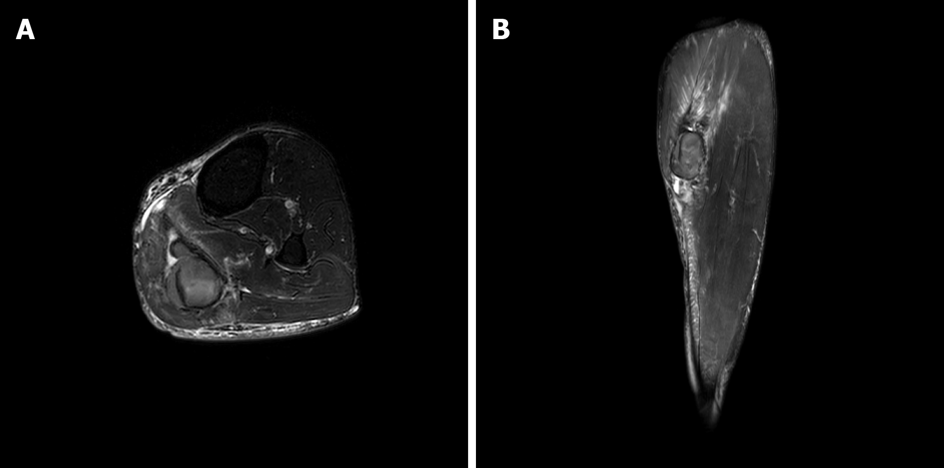
Initial diagnostic ultrasonography showed a hematoma in the left gastrocnemius muscle measuring 4.3 cm × 1.5 cm × 4.9 cm (Figure 1A and B). There was no active bleeding, and the hematoma was stiff and firm with low heterogeneity. Left tibial magnetic resonance imaging confirmed rupture of the medial head of the gastrocnemius muscle, with hematoma between the medial and lateral heads (Figure 2A and B).
The final diagnosis of the presented case is a painful hematoma in the left calf.
On the second hospital day, we applied ESWT to the hematoma for a total of 3000 shocks delivered at 6 Hz with 0.056 mJ/mm2. The shock waves were applied with a Dornier Aries® (Dornier MedTech Systems, Munich, Germany). We gradually increased the ESWT intensity.
After the first 1000 shocks, we moved the ankle passively and noted improved pain. After another 1000 shocks, the pain decreased further with passive dorsiflexion. Finally, after the final 1000 shocks, the patient was able to walk without crutches.
After the procedure, his pain decreased immediately to a VAS 3, and the mass softened. The hematoma measured 4.2 cm × 1.4 cm × 4.6 cm and its texture was more heterogeneous on ultrasonography compared to initial findings (Figure 3A-C). There were no adverse or unanticipated events.
The patient was discharged on the day he received ESWT with a prescription for Tramadol (Tridol®) for pain relief. Due to planned travel, he left overseas on the day of discharge. He remained abroad for three months and then returned to Korea. Upon return three months after the initial visit, he reported dramatic pain relief. He had experienced persistent discomfort in walking for about two weeks after treatment that resolved. But his pain did not recur. The hematoma gradually became smaller and finally resolved.
The current case describes successful treatment of a painful intramuscular hematoma with ESWT. The patient’s pain immediately decreased from VAS 7 to 3 after ESWT. In addition, the hematoma initially was stiff and firm with low heterogeneity on ultrasonography but became soft with greater heterogeneity after ESWT.
A hematoma can occur after muscle or soft tissue injury, when one or more blood vessels are injured and blood leaks under the dermis, into a joint, or between muscles. In general, hematomas break into fragments, are slowly absorbed by the body, and eventually evacuated via blood and lymph. However, this process can take time[8]. Järvinen et al[9] proposed that muscle strain injuries go through a three-phase healing process. Each of the steps in this process can be disrupted, which can lead to a chronic condition and failed repair, reflecting prolonged dysregulation and a maladaptive process that ultimately leads to tissue destruction[6]. Alessandrino et al[10] reported that most muscle lesions recover primarily through myofiber regeneration, but that healing after severe trauma or recurrence, occurs primarily through formation of a fibrotic scar[10]. A hematoma may not completely resolve when these anomalous processes continue. If this occurs, connective tissue can be deposited within the hematoma and calcium can be deposited in the tissue[11], which can cause pain and permanent poor mobility. This patient had a stiff, localized, oval hematoma with bruising on his left upper posterior calf. It had been two weeks since his injury, and these abnormal processes might have progressed since injury.
In this case, the initial diagnostic ultrasonography showed a firm, stiff hematoma with low heterogeneity in the left gastrocnemius muscle. Conforti described a potential complication of chronic organized hematoma[11]. Computed tomography shows the hematoma as a homogenous mass with capsule formation and a fibrous pseudo-capsule, whereas ultrasonography shows a multi-loculated cyst[11,12]. If the hematoma is surrounded by a fibrous capsule, it can harden, and cause persistent pain due to improper blood supply.
ESWT has been applied to various musculoskeletal conditions, including upper extremity conditions such as lateral epicondylitis and rotator cuff tendinopathy; it has also been applied to lower extremity conditions such as Achilles, patellar, and hamstring tendinopathies; as well as greater trochanteric pain syndrome[1]. ESWT is also used for non-union of long bone fractures, avascular necrosis of the femoral head, chronic diabetic and non-diabetic ulcers, and ischemic heart disease[2]. Table 1 shows the current applications of ESWT. In their narrative study, Reilly et al[1] proposed mechanisms of action for the shockwave as follows: Neovascularization at the tendon-bone junction, increased collagen synthesis and tissue remodeling, leukocyte infiltration, proliferation of tenocytes, mechanotransduction, stimulation of nociceptive C-fibers resulting in neuropeptide release, and nociceptor hyperstimulation. Wang[2] suggested that the majority of orthopedic shockwaves are used to induce microscopic interstitial and extracellular responses to tissue regeneration[2].
| Musculoskeletal disorders | Non-musculoskeletal disorders |
| Plantar fasciitis | Chronic diabetic foot ulcers |
| Achilles tendinopathy | Ischemic heart disease |
| Patellar tendinopathy | |
| Hamstring tendinopathy | |
| Greater trochanteric pain syndrome | |
| Medial tibial stress syndrome | |
| Non-union and delayed union of long bone fracture | |
| Avascular necrosis of the femoral head | |
| Stress fracture | |
| Lateral epicondylitis | |
| Tendinopathy of shoulder with or without calcification | |
| Peyronie’s disease | |
| Complex regional pain syndrome |
ESWT was first introduced into clinical practice in 1982 for urinary stone lithotripsy and was used for disintegrating renal stones or calcifications[1,2]. The mechanism of the ESWT therapeutic effect on shoulder calcification is uncertain. Rebuzzi et al[13] suggested that increasing stress within the therapeutic focus of the shockwave induces fragmentation and cavitation within amorphous calcifications, resulting in disorganization and disintegration of the deposite. The deposit may disappear as it breaks through into the adjacent vessels or surrounding soft tissue. Ogden et al[14] suggested that shock waves generate high stress forces that act on boundary interfaces and generate tensile forces that cause cavitation. According to their paper, the high pressure amplitude and the short rise time of the shock waves exceed the elastic strength of the stone, which cause the surface to disintegrate.
In this case, the pain had decreased since ESWT treatment, and we confirmed changes in heterogeneity on ultrasonography. We believe that the firm hematoma was softened by ESWT, resolving the firm mass effect that caused the pain.
There are several limitations to this report. First, the pain may have been reduced by another mechanism of ESWT. We suspected that the patient’s pain was relived because the mass softened after ESWT, which helped disorganize and disintegrate the deposit. However, other mechanisms of ESWT including gate-control theory, can affect pain relief. Therefore, further research may be required to confirm if pain is improved through other mechanisms. Second, there was no assessment to determine if the shockwave was strong enough to change the texture of the hematoma. Additional studies are needed to test the relationship between ESWT intensity and changes in hematoma texture. Finally, the pain relief could be attributable to medication. However, this is less likely because we confirmed a change in heterogeneity after treatment.
Hematoma is a possible complication of ESWT[2,15], and there are no case reports of ESWT as a therapeutic application for painful hematomas. However, as reviewed in Zissler et al[6], ESWT could reduce a chronic condition to an acute response. In addition, as mentioned above, ESWT may affect capsule breakdown and deposit disorganization in unabsorbable hematomas with fibrous capsules. Therefore, ESWT may be a new therapeutic approach for painful hematomas, and this case report may broaden the indications for ESWT and suggests new treatments for painful hematomas.
This is the first reported case of a painful intramuscular hematoma treated with ESWT. With ESWT treatment, the patient’s pain was immediately relieved, and the hematoma texture changed. We propose that painful hematoma could be a new indication for ESWT. Further investigation regarding the effects of ESWT and appropriate protocols for hematoma treatment are needed.
Manuscript source: Unsolicited manuscript
Specialty type: Rehabilitation
Country/Territory of origin: South Korea
Peer-review report’s scientific quality classification
Grade A (Excellent): 0
Grade B (Very good): B
Grade C (Good): 0
Grade D (Fair): 0
Grade E (Poor): 0
P-Reviewer: Isik A S-Editor: Zhang L L-Editor: A P-Editor: Li JH
| 1. | Reilly JM, Bluman E, Tenforde AS. Effect of Shockwave Treatment for Management of Upper and Lower Extremity Musculoskeletal Conditions: A Narrative Review. PM R. 2018;10:1385-1403. [RCA] [PubMed] [DOI] [Full Text] [Cited by in Crossref: 45] [Cited by in RCA: 66] [Article Influence: 9.4] [Reference Citation Analysis (0)] |
| 2. | Wang CJ. Extracorporeal shockwave therapy in musculoskeletal disorders. J Orthop Surg Res. 2012;7:11. [RCA] [PubMed] [DOI] [Full Text] [Full Text (PDF)] [Cited by in Crossref: 255] [Cited by in RCA: 310] [Article Influence: 23.8] [Reference Citation Analysis (0)] |
| 3. | Sakamoto A, Okamoto T, Matsuda S. Chronic Expanding Hematoma in the Extremities: A Clinical Problem of Adhesion to the Surrounding Tissues. Biomed Res Int. 2017;2017:4634350. [RCA] [PubMed] [DOI] [Full Text] [Full Text (PDF)] [Cited by in Crossref: 11] [Cited by in RCA: 18] [Article Influence: 2.3] [Reference Citation Analysis (0)] |
| 4. | Manenti G, Cavallo AU, Marsico S, Citraro D, Vasili E, Lacchè A, Forcina M, Ferlosio A, Rossi P, Floris R. Chronic expanding hematoma of the left flank mimicking a soft-tissue neoplasm. Radiol Case Rep. 2017;12:801-806. [RCA] [PubMed] [DOI] [Full Text] [Full Text (PDF)] [Cited by in Crossref: 6] [Cited by in RCA: 6] [Article Influence: 0.8] [Reference Citation Analysis (0)] |
| 5. | Reid JD, Kommareddi S, Lankerani M, Park MC. Chronic expanding hematomas. A clinicopathologic entity. JAMA. 1980;244:2441-2442. [PubMed] |
| 6. | Zissler A, Stoiber W, Pittner S, Sänger AM. Extracorporeal Shock Wave Therapy in Acute Injry care: A Systematic Review. Rehabilitation Process and Outcome. 2018. [RCA] [DOI] [Full Text] [Cited by in Crossref: 5] [Cited by in RCA: 5] [Article Influence: 0.7] [Reference Citation Analysis (0)] |
| 7. | Drüeke TB. A clinical approach to the uraemic patient with extraskeletal calcifications. Nephrol Dial Transplant. 1996;11 Suppl 3:37-42. [RCA] [PubMed] [DOI] [Full Text] [Cited by in Crossref: 44] [Cited by in RCA: 47] [Article Influence: 1.6] [Reference Citation Analysis (0)] |
| 8. | Negoro K, Uchida K, Yayama T, Kokubo Y, Baba H. Chronic expanding hematoma of the thigh. Joint Bone Spine. 2012;79:192-194. [RCA] [PubMed] [DOI] [Full Text] [Cited by in Crossref: 24] [Cited by in RCA: 27] [Article Influence: 1.9] [Reference Citation Analysis (0)] |
| 9. | Järvinen TA, Järvinen M, Kalimo H. Regeneration of injured skeletal muscle after the injury. Muscles Ligaments Tendons J. 2013;3:337-345. [PubMed] |
| 10. | Alessandrino F, Balconi G. Complications of muscle injuries. J Ultrasound. 2013;16:215-222. [RCA] [PubMed] [DOI] [Full Text] [Cited by in Crossref: 25] [Cited by in RCA: 30] [Article Influence: 2.5] [Reference Citation Analysis (0)] |
| 11. | Conforti M. The Treatment of Muscle Hematomas. In: Bisciotti G, Eirale C, ed. by. Muscle Injuries in Sport Medicine [Internet]. 2020 [cited 4 August 2020]. Available from: https://www.intechopen.com/books/muscle-injuries-in-sport-medicine/the-treatment-of-muscle-hematomas. |
| 12. | Nakano M, Kondoh T, Igarashi J, Kadowaki A, Arai E. A case of chronic expanding hematoma in the tensor fascia lata. Dermatol Online J. 2001;7:6. [PubMed] |
| 13. | Rebuzzi E, Coletti N, Schiavetti S, Giusto F. Arthroscopy surgery vs shock wave therapy for chronic calcifying tendinitis of the shoulder. J Orthop Traumatol. 2008;9:179-185. [RCA] [PubMed] [DOI] [Full Text] [Full Text (PDF)] [Cited by in Crossref: 37] [Cited by in RCA: 42] [Article Influence: 2.5] [Reference Citation Analysis (0)] |
| 14. | Ogden JA, Tóth-Kischkat A, Schultheiss R. Principles of shock wave therapy. Clin Orthop Relat Res. 2001: 8-17. [RCA] [PubMed] [DOI] [Full Text] [Cited by in Crossref: 290] [Cited by in RCA: 308] [Article Influence: 12.8] [Reference Citation Analysis (0)] |
| 15. | Kim H, Cheon JH, Lee DY, Cheon JH, Cho YK, Lee SH, Kang EY. Intramuscular Hematoma Following Radial Extracorporeal Shockwave Therapy for Chronic Neurogenic Heterotopic Ossification: A Case Report. Ann Rehabil Med. 2017;41:498-504. [RCA] [PubMed] [DOI] [Full Text] [Full Text (PDF)] [Cited by in Crossref: 4] [Cited by in RCA: 5] [Article Influence: 0.6] [Reference Citation Analysis (0)] |


