Published online Dec 26, 2020. doi: 10.12998/wjcc.v8.i24.6499
Peer-review started: September 23, 2020
First decision: October 18, 2020
Revised: October 30, 2020
Accepted: November 9, 2020
Article in press: November 9, 2020
Published online: December 26, 2020
Processing time: 87 Days and 10.6 Hours
Myiasis is a rare but risky pathology caused by a parasitic infestation of humans and animals by the dipterous larva. Oral myiasis occurs when soft tissues of the oral cavity are invaded by the larvae of flies. It is not a common disease for the reason that the oral cavity is not easily reachable for the fly to lay eggs. But it can cause pain, infection, uncomfortable feeling when the worms move, tissue destruction and/or even life-threatening hemorrhages.
We reported a case of oral myiasis after cerebral infarction in a 78-year-old male patient from southern China (Guangdong Zhanjiang). As a result of cerebral infarction, he suffered from right hemiplegia, mobility and mental decline for about 3 mo. He had difficulty swallowing and was fed via a feeding tube. He mostly engaged in mouth breathing and had poor oral and dental hygiene. More than 20 live larvae were collected from the patient’s oral cavity, which were localized in the maxillary gingiva, the mandibular gingiva and the tongue. The patient recovered after the routine oral cleaning, removal of maggots, debridement and anti-infection treatment.
Early diagnosis and treatment of this infestation are essential due to the bothersome symptoms, such as inflammation, intense anxiety over the larvae movement, possible serious complications, etc. Clinical staff should be familiar with this infestation, and this disease should be considered, especially in physically and mentally disabled patients or those at significant risk for infection. Necessary measures, including good sanitation, personal and environmental hygiene and special care should be adopted so as to prevent this disease.
Core Tip: We reported a case of oral myiasis after cerebral infarction in a 78-year-old male patient from southern China. He suffered from right hemiplegia, mobility and mental decline for about 3 mo. He was fed via a feeding tube and mostly engaged in mouth breathing with poor oral and dental hygiene. More than 20 live larvae were collected from the patient’s oral cavity. The patient recovered after the routine oral cleaning, removal of maggots, debridement and anti-infection treatment. Early diagnosis and treatment of this infestation are essential and clinical staff should be familiar with this infestation.
- Citation: Zhang TZ, Jiang Y, Luo XT, Ling R, Wang JW. Oral myiasis after cerebral infarction in an elderly male patient from southern China: A case report. World J Clin Cases 2020; 8(24): 6499-6503
- URL: https://www.wjgnet.com/2307-8960/full/v8/i24/6499.htm
- DOI: https://dx.doi.org/10.12998/wjcc.v8.i24.6499
The term myiasis was first introduced in 1840 to describe a parasitic infestation of humans and animals by dipterous larva. Myiasis can affect the skin, external orifices (eyes, ears, nasal cavities, oral cavity, vagina and anus), and internal organs (intestines and urinary tract)[1]. Oral myiasis occurs when the larvae of flies invade soft tissues of the oral cavity. It is a rare pathology because the oral cavity is not easily reachable for the fly to lay eggs. Oral myiasis can cause pain, infection, uncomfortable feeling when the worms move and tissue destruction. It can even destroy vital tissues, which may induce severe or life-threatening hemorrhages, thus posing a serious risk to the patient’s life[2,3].
Poor oral hygiene, halitosis, facial trauma, suppurative lesions, ulcerative lesions, wound extraction, fumigating cancers, senility, immunocompromised state, unhygienic living conditions, learning disabilities, neurological deficit and other physically and mentally challenging conditions have all been associated with oral myiasis[4,5].
Myiasis is prevalent in tropical and subtropical areas. Zhanjiang city is situated in the Guangdong province of southern China, which is a subtropical area. Herein, we reported a rare case of oral myiasis after cerebral infarction in a 78-year-old male patient from Zhanjiang.
On December 6, 2019, a 78-year-old male patient was admitted to The Department of Stomatology, The Affiliated Hospital of Guangdong Medical University due to the infection and erosion of the gum, suppuration of the tongue and the discovery of maggot-like worms.
Two weeks ago, family members of the patient noticed the infection and erosion of the gum and suppuration of the tongue. The maggot-like worms were observed the past 3 d.
The patient’s general health was poor. In September 2019, he was admitted to The People’s Hospital of Maoming (a local hospital near his house) due to cerebral infarction, for which he was treated with left middle cerebral artery balloon dilation, artery antithrombotic, thrombus aspiration and stent implantation. Right side catheterization of the internal jugular vein was also applied. Nonetheless, as a result of the cerebral infarction, he still suffered from right hemiplegia, mobility and mental decline. He had difficulty swallowing and was fed via a feeding tube.
The histories of diabetes, hypertension and other chronic diseases were denied. The histories of hepatitis, tuberculosis and other infectious diseases, trauma, blood transfusion and allergy were denied too. The vaccination history was unknown.
Physical examination revealed right hemiplegia, mental decline and inability to communicate. Body temperature was 38.5 °C; pulse was 137 times/min, breath was 20 times/min, and blood pressure was 128/72 mmHg.
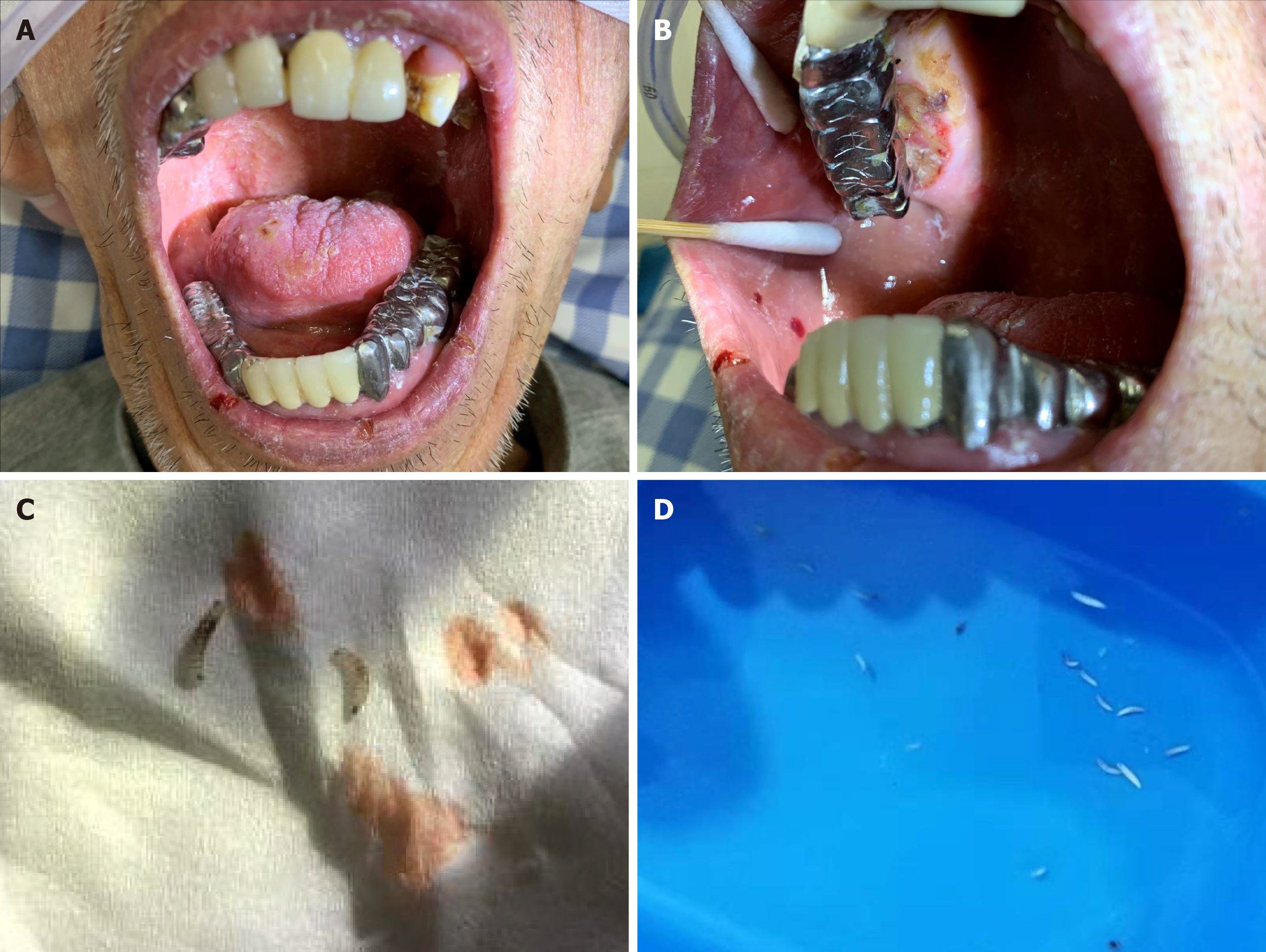
The oral and maxillofacial regions were symmetrical. The feeding tube was in place. Swallowing was difficult. The patient mostly relied on mouth breathing. He had poor oral and dental hygiene, halitosis, a swollen tongue with a smooth back and atrophic tongue papillae. The duct openings of the bilateral submandibular glands were red and swollen. Fixed bridge repair of the right maxillary and bilateral mandibular teeth was observed. The crown edges were not fitted. The degree of teeth mobility was I-II. The palatal gum of the right maxillary teeth was erosive and ulcerated and about the size of a “soybean”. Pus overflowed when squeezed, and on the buccal gum, a white maggot about 1.0 cm long was seen.
There was a fistula about 0.5 cm ×0.5 cm in size in the front of the right tongue. Pus overflowed with maggots wriggling in the fistula.
The patient had more than 20 maggots in his oral cavity in total. All of them were milky white and cylindrical with sharp front and blunt back. Peristalsis was obvious as well as folds in the body (Figure 1).
Leukocyte count was 3.45 × 10⁹/L. Neutrophil ratio was increased at 89.25%, Lymphocyte ratio and monocyte ratio were decreased at 9.28% and 1.35%, respectively. The rest of the routine laboratory examinations were normal.
Computed tomography scan showed many artifacts in the oral cavity and the adjacent structures were not clear. Irregular destruction in the left region of the maxilla bones was revealed, considering the possibility of inflammatory lesions. Osteoporosis of the maxilla and mandible bones was also observed. The soft tissue around the upper alveolar was swollen with a little gas accumulated. The mucosa of the bilateral ethmoid sinus and maxillary sinus was slightly thickened.
Based on all of the observations above, the case was diagnosed as oral myiasis.
Treatment was carried out to resolve the infection of the mouth and the maggots. Local application of iodoform for a minimum of 20-30 min was used to irritate the maggots and force them out of their hiding places. Maggots were also manually removed with blunt tweezers and curved forceps. The patient’s mouth was thoroughly irrigated three times a day with a solution of 3% hydrogen peroxide, normal saline, 2% sodium bicarbonate and 0.2% chlorhexidine mouth wash. The rotten tissues were manually removed with tweezers and curved forceps. Endovenous rehydration was performed. The mouth was covered with wet gauze to prevent dry mouth and avoid the contact of the mouth with flies or their eggs.
After 7 d of hospitalization, the patient recovered well. His general condition was stable. No maggots were found in the mouth, and there was no obvious acute inflammation in his oral cavity. No evident hyperemia or swelling of gingiva and tongue were found. The ulcerated gum and the fistula of the tongue were healed. The patient was discharged on December 13, 2019. Family members were advised to maintain the patient’s oral hygiene and prevent contact with flies. A head cap was suggested to prevent the patient from keeping his mouth open for a long time and prevent temporomandibular joint related dislocations.
A follow-up visit was suggested 1 mo later; however, the patient did not return due to family members’ personal reasons. Upon calling, we were told the patient was in good condition. We suggested re-examination once every 3 mo.
Myiasis is prevalent in tropical and subtropical areas. In China, myiasis often occurs in southern China, which is characterized by a hot and humid climate. This is favorable for larvae growth. Zhanjiang city of Guangdong province is situated in southern China and in the subtropical area.
Most of the reported myiasis tend to be ocular myiasis and dermato-myiasis[1]. Oral myiasis is very rarely observed. In this case, the patient’s general health was poor, and he had a history of cerebral infarction. When he was brought to our hospital, he suffered from hemiplegia on the right side of the body, inconvenient movement and mental retardation. Consequently, the patient was in no condition to engage in general and oral health care. Also, there were many bad restorations in his mouth, which led to poor oral hygiene and obvious inflammation, suppuration and erosion of gums and mucous membranes. The oral environment was suitable for the larvae growth. The patient tended to engage in mouth breathing exclusively. Because flies may lay eggs or larvae near the mouth and nose, when the patient deeply inhaled, he would also inhale the eggs or larvae into the mouth, which then developed into maggots leading to the onset of the disease.
Treatment in this study included the local application of iodoform, routine oral cleaning, removal of maggots, debridement, and anti-infection therapy. We locally used iodoform to irritate the maggots and force them out of their hiding places. Other substances such as turpentine oil, mineral oil, olive oil, chloroform, creosote, phenol and calomel can also be used[4]. Unfortunately, in the present case, the patient’s bad denture was not restored because the patient could not cooperate with doctors. His relatives also thought it was unnecessary as he could not swallow or chew food and was fed via a feeding tube.
Early diagnosis and treatment of this infestation are essential due to the bothersome symptoms such as inflammation, intense anxiety over the larvae movement, possible serious complications, etc[2,3]. Clinical staff should be familiar with this infestation, and this parasite should be considered, especially in risky patients. Necessary measures, including good sanitation, personal and environmental hygiene and special care should be adopted to prevent this disease[4,5].
The authors acknowledge the help of all the co-workers.
Manuscript source: Unsolicited manuscript
Specialty type: Medicine, research and experimental
Country/Territory of origin: China
Peer-review report’s scientific quality classification
Grade A (Excellent): 0
Grade B (Very good): 0
Grade C (Good): C
Grade D (Fair): 0
Grade E (Poor): 0
P-Reviewer: Treviño González JL S-Editor: Huang P L-Editor: Filipodia P-Editor: Li JH
| 1. | Francesconi F, Lupi O. Myiasis. Clin Microbiol Rev. 2012;25:79-105. [RCA] [PubMed] [DOI] [Full Text] [Cited by in Crossref: 335] [Cited by in RCA: 322] [Article Influence: 24.8] [Reference Citation Analysis (0)] |
| 2. | Taş Cengiz Z, Yılmaz H, Beyhan YE, Yakan Ü, Ekici A. An Oral Myiasis Case Caused by Diptera (Calliphoridae) Larvae in Turkey. Turkiye Parazitol Derg. 2019;213-215. [RCA] [PubMed] [DOI] [Full Text] [Cited by in Crossref: 3] [Cited by in RCA: 3] [Article Influence: 0.6] [Reference Citation Analysis (0)] |
| 3. | Ahmadpour E, Youssefi MR, Nazari M, Hosseini SA, Rakhshanpour A, Rahimi MT. Nosocomial Myiasis in an Intensive Care Unit (ICU): A Case Report. Iran J Public Health. 2019;48:1165-1168. [PubMed] |
| 4. | Bhansali SP, Tiwari AD, Gupta DK, Bhansali S. Oral myiasis in paralytic patients with special needs: A report of three cases. Natl J Maxillofac Surg. 2018;9:110-112. [RCA] [PubMed] [DOI] [Full Text] [Full Text (PDF)] [Cited by in Crossref: 2] [Cited by in RCA: 2] [Article Influence: 0.3] [Reference Citation Analysis (1)] |


