Published online Dec 26, 2020. doi: 10.12998/wjcc.v8.i24.6450
Peer-review started: August 12, 2020
First decision: August 21, 2020
Revised: August 31, 2020
Accepted: September 25, 2020
Article in press: September 25, 2020
Published online: December 26, 2020
Processing time: 129 Days and 4.7 Hours
Scapular fracture has a low incidence rate, accounting for 0.4%-0.9% of all fractures, and scapular neck fractures are extremely rare, comprising approximately 7%-25% of all scapular fractures. Scapular neck fractures are often studied as case reports mostly accompanied by other injuries, thus leading to confusion. All previous cases of scapular neck fractures are not associated with rotator cuff injuries.
A 62-year-old man was admitted to our emergency department 6 h after his right shoulder and back were impacted by heavy objects. The patient presented chest tightness and shortness of breath. Chest computed tomography (CT) showed pneumohemothorax, multiple rib fractures, and right scapula fractures. Three-dimensional CT reconstruction of the right shoulder joint showed a trans-spinous scapular neck fracture with a glenohumeral joint dislocation. Rotator cuff injury was suspected because the patient had a glenohumeral joint dislocation and was then confirmed by shoulder magnetic resonance imaging. A staged surgery was performed, including open reduction and internal fixation of the right scapula fracture and repairing of rotator cuff by right shoulder arthroscopy. At the 5-mo follow-up, the fracture line was blurred and the shoulder joint function was good.
Fracture of the scapular neck combined with rotator cuff tear is rare and the rotator cuff injury should not be ignored in clinical work. Stable internal fixation combined with secondary arthroscopic repair of rotator cuff tear can achieve good results.
Core Tip: We describe a patient diagnosed with fracture of the scapular neck combined with rotator cuff tear. Scapular fracture has a low incidence rate and scapular neck fractures are extremely rare. Fracture of the scapular neck combined with rotator cuff tear has not been reported previously. Rotator cuff tear should not be ignored in clinical work when treating this type of fracture. Stable internal fixation combined with secondary arthroscopic repair of rotator cuff tear can achieve good results.
- Citation: Chen L, Liu CL, Wu P. Fracture of the scapular neck combined with rotator cuff tear: A case report. World J Clin Cases 2020; 8(24): 6450-6455
- URL: https://www.wjgnet.com/2307-8960/full/v8/i24/6450.htm
- DOI: https://dx.doi.org/10.12998/wjcc.v8.i24.6450
Fractures of the scapula are relatively rare and account for less than 1% of all fractures and 3%-5% of shoulder girdle fractures[1]. Most scapular fractures occur in the body, and acceptable results from conservative treatment have been achieved. Fractures of the scapula neck are rare, accounting for approximately 7%-25% of all scapula fractures[2-4], and is accompanied by complex anatomical structures, confusion in diagnosis, and controversial treatment[4-7]. Bartoníček et al[3,8] summarized reports and added their own cases to describe the diagnosis, classification, and treatment of scapular neck fractures and recommended surgical treatment for displaced scapular neck fractures.
All previous cases of scapular neck fractures are not associated with rotator cuff injuries. Here, we report an extremely rare scapular neck case with rotator cuff injury and biceps interposition, analyze the injury mechanism, present radiographic images, and describe the treatment and follow-up procedures.
A 62-year-old man was admitted to our emergency department 6 h after his right shoulder and back were impacted by heavy objects.
The patient presented chest tightness and shortness of breath. Chest computed tomography (CT) was performed in the emergency department. He experienced pneumohemothorax, multiple rib fractures, and right scapula fractures. Hence, thoracic closed drainage was performed, and his right hand was suspended by a sling. After the patient’s vital signs became stable, he was transferred to our department.
The patient denied any previous medical history of the right shoulder and surgery.
Physical examination revealed tenderness in the right shoulder, limited movement of the right shoulder, and no numbness, limitation of finger movement, or signs of vascular injury.
His hemoglobin was 98 g/L.
Given the confirmed right scapular fracture by previous emergency CT, no right shoulder X-ray was performed. Three-dimensional (3D) CT reconstruction of the right shoulder joint (Figure 1) showed a trans-spinous scapular neck fracture with a glenohumeral joint dislocation.
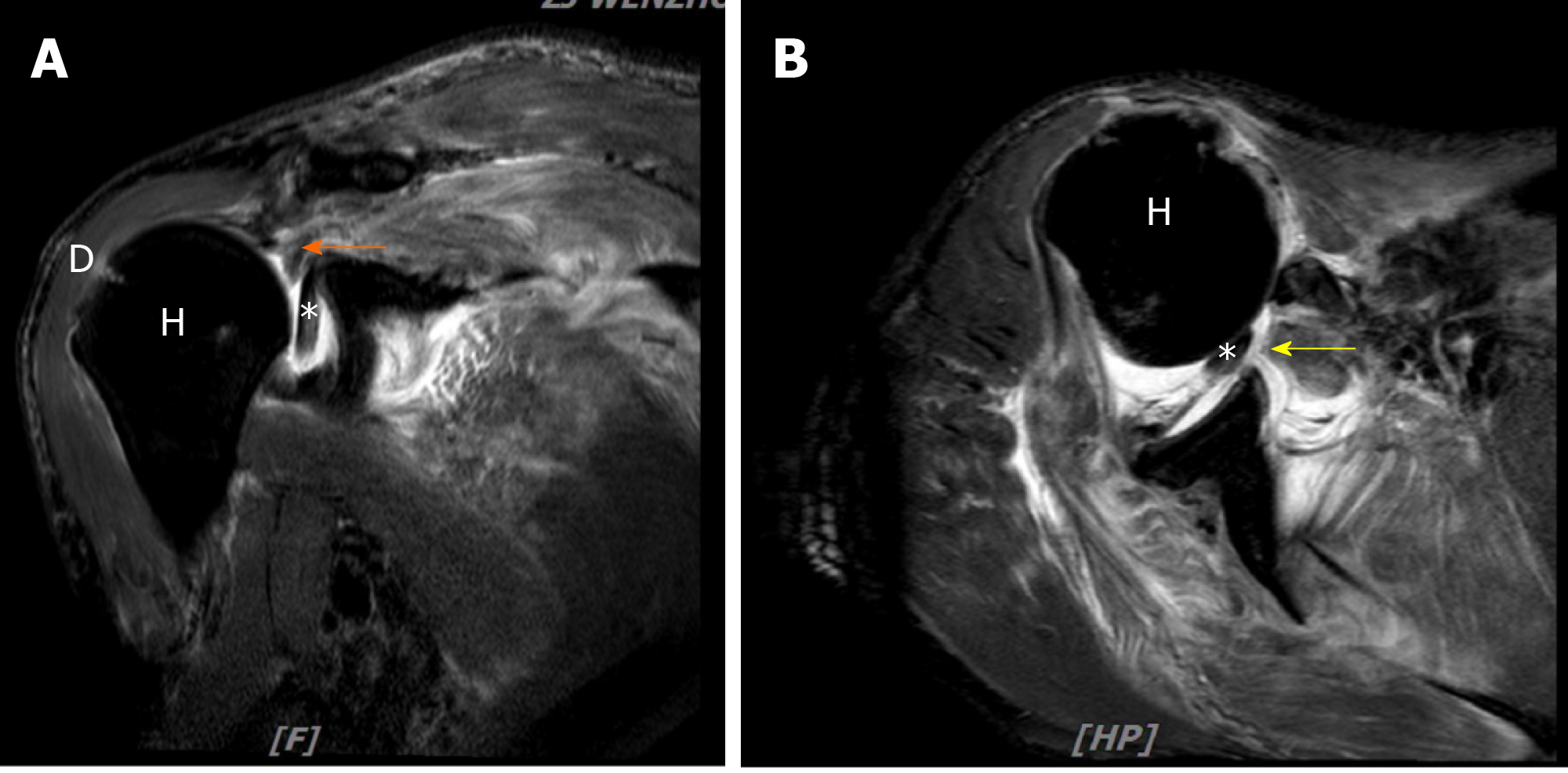
Rotator cuff injury was suspected because the patient had a glenohumeral joint dislocation. Hence, magnetic resonance imaging (MRI) of the right shoulder was performed before surgery (Figure 2), which showed full-thickness tears of the supraspinatus, subscapularis tendons off their respective footprints, and the tendon of long head biceps incarcerated in the glenohumeral joint.
Based on the history and preoperative imaging examination, this patient was diagnosed with fracture of the scapular neck combined with rotator cuff tear.
We made a staged surgical plan. The patient underwent open reduction and internal fixation of the right scapula fracture 13 d after the injury. After receiving general anesthesia, the patient was placed in the left semi-prone position, and the right upper limb was abducted at 90°. The scapula was approached by an L-shaped Judet incision, which distally extended from the posterior edge of acromion along the spine and curved along the medial scapular border to the inferior angle. Periosteocutaneous flaps were raised, and the infraspinatus, teres minor, and deltoid muscles were elevated from posterior scapula body to subsequently expose the scapular spine and body. The scapula spine was severely comminuted. A 3.5 mm locking plate was applied, and a 2.7 mm reconstruction plate was used to enhance the fixation strength. The lateral border of the scapula was fixed with two 2.7 mm reconstruction plates. Postoperative X-ray and CT showed that anatomical reduction was achieved, and the internal fixation position was stable. However, the glenohumeral joint was still dislocated (Figure 3).
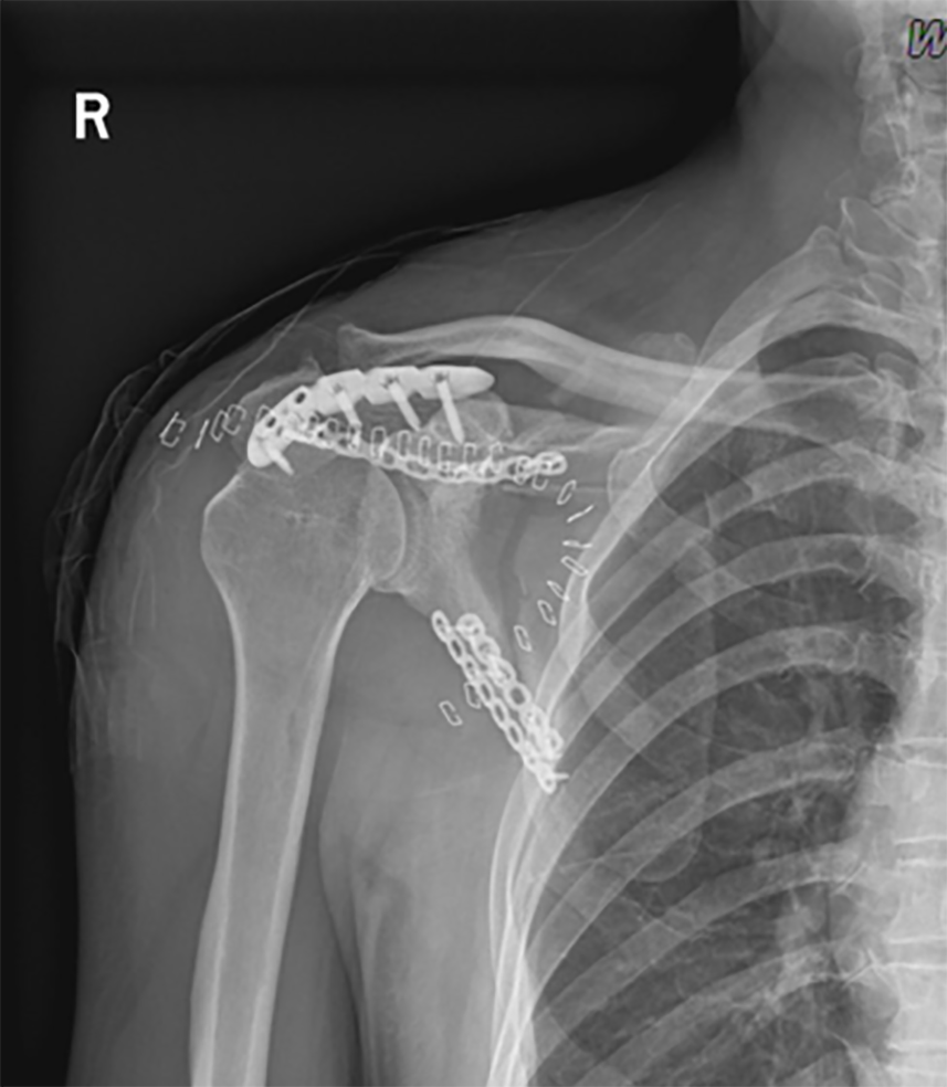
Five days after the first stage operation, the patient underwent right shoulder arthroscopy under general anesthesia. Intraoperative investigations confirmed a full-thickness tear of the supraspinatus tendon, subscapularis tendons off their respective footprints, and incarcerated tendon of long head biceps in the glenohumeral joint. The bicep long head tendon was cut and re-fixed in the intertubercular sulcus, and the rotator cuff was repaired with an anchor screw. Postoperative X-ray confirmed concentric reduction of the humeral head within the glenoid concavity (Figure 4).
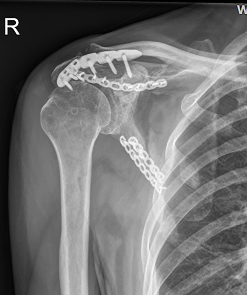
The patient started passive functional exercise immediately after the operation. Active functional exercise was allowed 6 wk post-operation. At the 5-mo follow-up, a small translucent shadow was observed on the lateral margin, and the fracture line was blurred (Figure 5). The shoulder joint activity was completely non-painful, and the function was good. Constant score was 90.
Scapular fractures have a low incidence rate, and scapular neck fractures are even rare, accounting for approximately 7%-25% of all scapular fractures[2-4]. The difference in the incidence rate may be due to the confusion of diagnosis and classification[3] as most scapular neck fractures are diagnosed and classified by X-ray[2,4,9-11] and subject to conservative treatment. Hence, these cases cannot be verified intra-operatively. For the diagnosis and classification of fractures with such complex shapes, X-ray is insufficient and will lead to misdiagnosis. CT with 3D reconstruction is necessary for the accurate diagnosis and classification of scapular fractures[8,12,13]. Bartoníček et al[3] treated and analyzed 17 cases of scapular neck fractures and concluded that scapular fractures can be summarized into three types, namely, anatomical, surgical, and trans-spinous neck fractures. Our case belongs to a trans-spinous neck fracture. However, none of the previously reported scapular neck fracture cases is combined with rotator cuff injury. To our knowledge, this study is the first reported case of scapular neck fracture with rotator cuff injury.
The injury mechanism of trans-spinous scapular neck fracture is high-energy trauma that directly hits the scapula from behind, thus causing the breakage of both pillars of the scapula, including the lateral border of the scapular body and the scapular spine[3]. In our case, the patient also hit his right back and chest with a falling heavy object. This condition can also be verified from the patient's chief complaint and accompanying pneumothorax and multiple rib fractures. We speculate that the energy continued to act upon the proximal humerus, further tearing the rotator cuff and leading to the long bicep tendon being stuck into the glenohumeral joint. The upward pulling force of the deltoid muscle caused the dislocation of the glenohumeral joint[14,15]. This type of fracture is characterized by the comminution of the lateral border of scapular body and intercalcar fragments in the infraspinous fossa. Therefore, Bartoníček et al[3] speculated that this fracture type is a pre-stage of comminuted fractures of the scapular body. The tear of traumatic rotator cuff is typically large and involves the subscapularis[16]. Our case presents the characteristics of trans-spinous scapular neck fractures and rotator cuff tear as reported in the literature.
With the growing understanding of scapular neck fractures and the development of internal fixation, this type of fracture has become increasingly inclined to surgery from the previous conservative treatment. The stable internal fixation is the basis for early functional exercise and reducing complications[3], and our choice of scapula spine and lateral border double-steel plate fixation technology provides sufficient fixation strength. Strong surgical indications were observed for traumatic rotator cuff tear, and arthroscopic repair is the first choice[17]. The anatomical reduction of the scapula with internal fixation at the first stage operation provides the cornerstone for the soft tissue repair of the rotator cuff at the second stage.
Rotator cuff tears are common in shoulder dislocations and fractures of the proximal humerus, and this condition has received attention. However, scapular fractures are not commonly accompanied by rotator cuff tear, especially, by biceps interposition simultaneously. Wyatt et al[15] reported a rare anterior superior dislocation of the humeral head and found that this dislocation pattern requires a unique combination of injuries, including a massive rotator cuff injury that resulted in the mobilization of the humeral head and a lack of a superior boundary and the incarceration of bicep tendon in the glenohumeral joint. The rotator cuff tear characteristics and dislocation in our case are identical to their case. In clinical practice, when we encounter patients with scapular neck fractures, we must not ignore the possibility of soft tissue damage. With this unique humeral head dislocation, we must consider rotator cuff tear and biceps interposition. And the necessity of taking MRI should be suggested in case of soft tissue damage.
We generally achieved satisfactory results for this rare case of scapular neck fracture with rotator cuff tear and biceps interposition through stable internal fixation combined with secondary arthroscopic repair of rotator cuff tear. Scapular neck fracture may be accompanied by rotator cuff injury, which should not be ignored in clinical work.
Fracture of the scapular neck combined with rotator cuff tear is rare. In clinical practice, when we encounter patients with scapular neck fractures, especially with this unique humeral head dislocation, we must consider rotator cuff injury. MRI is essential for diagnosis. Stable internal fixation combined with secondary arthroscopic repair of rotator cuff tear can achieve good results.
We thank the patient who participated in this study.
Manuscript source: Unsolicited manuscript
Specialty type: Medicine, research and experimental
Country/Territory of origin: China
Peer-review report’s scientific quality classification
Grade A (Excellent): 0
Grade B (Very good): B, B
Grade C (Good): 0
Grade D (Fair): D, D
Grade E (Poor): 0
P-Reviewer: Park JB, Thanindratarn P S-Editor: Yan JP L-Editor: Wang TQ P-Editor: Xing YX
| 1. | Rowe CR. Fractures of the scapula. Surg Clin North Am. 1963;43:1565-1571. [RCA] [PubMed] [DOI] [Full Text] [Cited by in Crossref: 88] [Cited by in RCA: 72] [Article Influence: 2.5] [Reference Citation Analysis (0)] |
| 2. | Ada JR, Miller ME. Scapular fractures. Analysis of 113 cases. Clin Orthop Relat Res. 1991;(269):174-180. [RCA] [PubMed] [DOI] [Full Text] [Cited by in Crossref: 7] [Cited by in RCA: 6] [Article Influence: 0.2] [Reference Citation Analysis (0)] |
| 3. | Bartoníček J, Tuček M, Frič V, Obruba P. Fractures of the scapular neck: diagnosis, classifications and treatment. Int Orthop. 2014;38:2163-2173. [RCA] [PubMed] [DOI] [Full Text] [Cited by in Crossref: 22] [Cited by in RCA: 19] [Article Influence: 1.7] [Reference Citation Analysis (0)] |
| 4. | Goss TP. Fractures of the glenoid neck. J Shoulder Elbow Surg. 1994;3:42-52. [RCA] [PubMed] [DOI] [Full Text] [Cited by in Crossref: 41] [Cited by in RCA: 23] [Article Influence: 0.7] [Reference Citation Analysis (0)] |
| 5. | de Beer JF, Berghs BM, van Rooyen KS, du Toit DF. Displaced scapular neck fracture: a case report. J Shoulder Elbow Surg. 2004;13:123-125. [RCA] [PubMed] [DOI] [Full Text] [Cited by in Crossref: 7] [Cited by in RCA: 6] [Article Influence: 0.3] [Reference Citation Analysis (0)] |
| 6. | Bozkurt M, Can F, Kirdemir V, Erden Z, Demirkale I, Başbozkurt M. Conservative treatment of scapular neck fracture: the effect of stability and glenopolar angle on clinical outcome. Injury. 2005;36:1176-1181. [RCA] [PubMed] [DOI] [Full Text] [Cited by in Crossref: 76] [Cited by in RCA: 59] [Article Influence: 3.0] [Reference Citation Analysis (0)] |
| 7. | van Noort A, van Kampen A. Fractures of the scapula surgical neck: outcome after conservative treatment in 13 cases. Arch Orthop Trauma Surg. 2005;125:696-700. [RCA] [PubMed] [DOI] [Full Text] [Cited by in Crossref: 43] [Cited by in RCA: 45] [Article Influence: 2.3] [Reference Citation Analysis (0)] |
| 8. | Bartoníček J, Frič V, Tuček M. Fractures of the anatomical neck of the scapula: two cases and review of the literature. Arch Orthop Trauma Surg. 2013;133:1115-1119. [RCA] [PubMed] [DOI] [Full Text] [Cited by in Crossref: 10] [Cited by in RCA: 11] [Article Influence: 0.9] [Reference Citation Analysis (0)] |
| 9. | Hitzrot JM, Bolling RW. FRACTURES OF THE NECK OF THE SCAPULA. Ann Surg. 1916;63:215-236. [RCA] [PubMed] [DOI] [Full Text] [Cited by in Crossref: 15] [Cited by in RCA: 15] [Article Influence: 0.8] [Reference Citation Analysis (0)] |
| 10. | Arts V, Louette L. Scapular neck fractures; an update of the concept of floating shoulder. Injury. 1999;30:146-148. [RCA] [PubMed] [DOI] [Full Text] [Cited by in Crossref: 31] [Cited by in RCA: 20] [Article Influence: 0.8] [Reference Citation Analysis (0)] |
| 11. | Pace AM, Stuart R, Brownlow H. Outcome of glenoid neck fractures. J Shoulder Elbow Surg. 2005;14:585-590. [RCA] [PubMed] [DOI] [Full Text] [Cited by in Crossref: 58] [Cited by in RCA: 43] [Article Influence: 2.2] [Reference Citation Analysis (0)] |
| 12. | Bartoníček J, Frič V. Scapular body fractures: results of operative treatment. Int Orthop. 2011;35:747-753. [RCA] [PubMed] [DOI] [Full Text] [Cited by in Crossref: 46] [Cited by in RCA: 43] [Article Influence: 2.9] [Reference Citation Analysis (0)] |
| 13. | Cole PA, Freeman G, Dubin JR. Scapula fractures. Curr Rev Musculoskelet Med. 2013;6:79-87. [RCA] [PubMed] [DOI] [Full Text] [Cited by in Crossref: 82] [Cited by in RCA: 67] [Article Influence: 5.6] [Reference Citation Analysis (0)] |
| 14. | Plachel F, Korn G, Abdic S, Moroder P. Acute locked superior shoulder dislocation in a patient with cuff tear arthropathy. BMJ Case Rep. 2018;2018:bcr2018225237. [RCA] [PubMed] [DOI] [Full Text] [Cited by in Crossref: 3] [Cited by in RCA: 5] [Article Influence: 0.7] [Reference Citation Analysis (0)] |
| 15. | Wyatt AR 2nd, Porrino J, Shah S, Hsu JE. Irreducible superolateral dislocation of the glenohumeral joint. Skeletal Radiol. 2015;44:1387-1391. [RCA] [PubMed] [DOI] [Full Text] [Cited by in Crossref: 6] [Cited by in RCA: 8] [Article Influence: 0.8] [Reference Citation Analysis (0)] |
| 16. | Mall NA, Lee AS, Chahal J, Sherman SL, Romeo AA, Verma NN, Cole BJ. An evidenced-based examination of the epidemiology and outcomes of traumatic rotator cuff tears. Arthroscopy. 2013;29:366-376. [RCA] [PubMed] [DOI] [Full Text] [Cited by in Crossref: 104] [Cited by in RCA: 107] [Article Influence: 8.9] [Reference Citation Analysis (0)] |
| 17. | Haviv B, Rutenberg TF, Bronak S, Yassin M. Arthroscopic rotator cuff surgery following shoulder trauma improves outcome despite additional pathologies and slow recovery. Knee Surg Sports Traumatol Arthrosc. 2018;26:3804-3809. [RCA] [PubMed] [DOI] [Full Text] [Cited by in Crossref: 8] [Cited by in RCA: 6] [Article Influence: 0.9] [Reference Citation Analysis (0)] |


